Introduction
Mpox, historically known as monkeypox, is a zoonotic viral disease caused by mpox virus, a double-stranded DNA virus in the genus
Orthopoxvirus and family Poxviridae [
1,
2]. The virus is closely related to variola virus, the causative agent of smallpox [
1]. Traditionally, 2 major clades of mpox virus have been recognized: the West African clade and the Congo Basin clade. The West African clade has been associated with a milder disease course, less frequent human-to-human transmission, and lower mortality than the Congo Basin clade [
1–
3].
On 23 July 2022, the World Health Organization (WHO) declared the multinational mpox outbreak a Public Health Emergency of International Concern (PHEIC). The outbreak was considered atypical because many cases occurred in countries without established endemic transmission and had no epidemiological link to historically endemic areas. Transmission predominantly affected men who have sex with men (MSM), although other populations were also affected [
4–
7]. The PHEIC designation was lifted on May 10, 2023 [
4]. Reported cases were found to have originated from the milder West African clade [
3,
5].
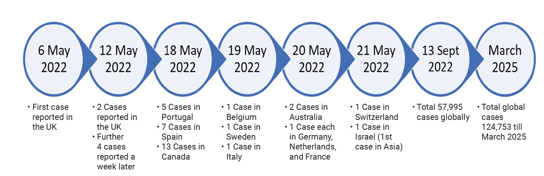
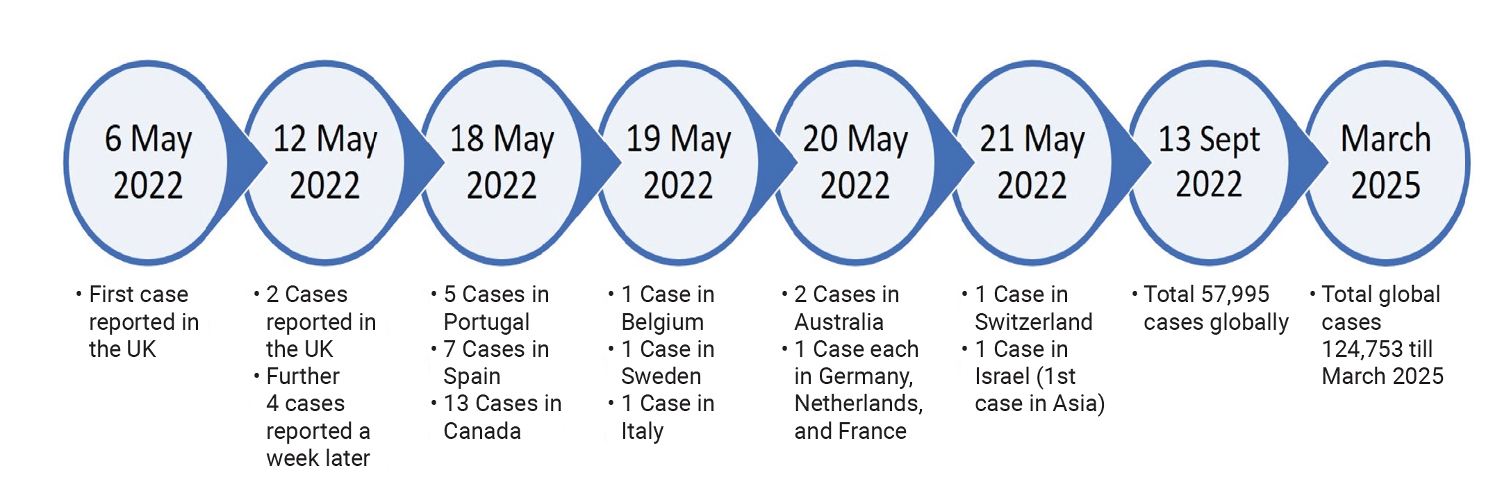
The first European case was identified in the United Kingdom on May 6, 2022 in an individual who had returned from Nigeria [
3,
8]. Additional cases were reported in the United Kingdom, the United States, Spain, Canada, Belgium, Sweden, Italy, France, Portugal, Germany, the Netherlands, Switzerland, Israel, and Australia in May 2022. Some cases had no known links to one another or to endemic areas. By September 13, 2022, nearly 58,000 confirmed cases had been reported in more than 100 countries, including 20,733 cases in the United States alone [
8,
9]. As of August 2023, 80,000 cases of atypical mpox had been reported [
9]. In India, 30 cases had been reported by September 2024 after the PHEIC declaration [
10].
Differences from Classical Mpox
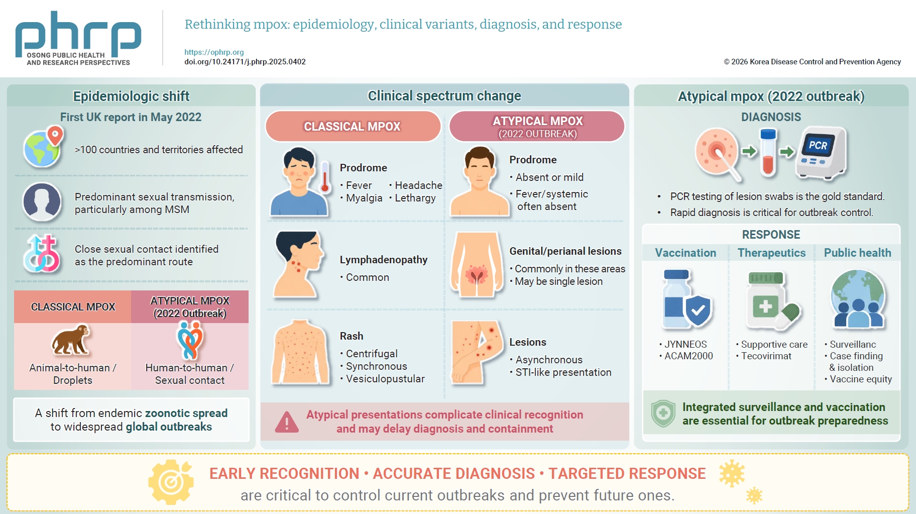
Classical disease begins with a prodrome of fever, lethargy, and lymphadenopathy, followed by a characteristic vesiculopustular rash [
11,
12]. The clinical manifestations of atypical mpox may differ from classical presentations. Although the characteristic rash and lesions are still present, atypical cases have shown a higher prevalence of genital and perianal lesions, reported in 73% of cases, as well as a higher incidence of proctitis [
6]. Single genital ulcers have also been reported in some cases [
13]. Several cases have been asymptomatic or mild [
3]. Prodromal symptoms may be absent in some cases and prolonged in others [
6]. Several case reports have also described atypical mpox in patients with human immunodeficiency virus (HIV) infection. Oral and tonsillar ulcers, epiglottitis, pharyngitis, and dysphagia are common oropharyngeal manifestations [
13]. Cutaneous lesions are mainly vesiculopustular (58%), although macular, pustular, vesicular, crusted, and multiphasic lesions have also been observed [
4].
The emergence of atypical mpox raised substantial global public health concerns. Its distinct epidemiological and clinical features challenged existing knowledge and preparedness efforts. As cases increased in non-endemic regions, understanding the characteristics, transmission dynamics, and potential impact of atypical mpox became increasingly important. This review provides a comprehensive overview of current knowledge and research on atypical mpox, including contemporary trends in clinical presentation, to reduce unfamiliarity and improve clinician awareness.
Epidemiological Aspects
Endemic, or typical, mpox is transmitted primarily through large respiratory droplets, followed by fomites and close physical contact with cases or infected animals [
3]. In atypical mpox, human-to-human transmission via respiratory droplets is not considered a major route. Early epidemiological data indicated that the atypical mpox outbreak primarily affected MSM, who accounted for approximately 98% of cases, particularly those with multiple or anonymous sexual partners [
3,
4,
14]. However, as the outbreak progressed, cases were also reported in other population groups, including women and children, with evidence suggesting a higher risk of severe disease in these subgroups [
15]. A total of 41% of patients were HIV-positive, but most did not have active immunodeficiency and had low viral loads [
4]. These data suggest that HIV positivity is better regarded as a coexisting condition than as an independent risk factor. However, HIV infection could be an important comorbidity in patients with immunodeficiency and may influence disease severity.
Progression of the Pandemic
Countries with endemic mpox virus transmission include Cameroon, Sierra Leone, Liberia, Côte d’Ivoire, Benin, Nigeria, Gabon, the Republic of the Congo, the Central African Republic, South Sudan, and the Democratic Republic of the Congo [
3,
8]. However, the course of atypical mpox has been erratic. Unlike classical mpox, atypical mpox has been estimated to include asymptomatic cases [
3]. Such cases may have been present in endemic regions before the 2022 outbreak. The first European case, involving a man who had traveled from Nigeria to the United Kingdom, was detected on May 6, 2022. The virus subsequently spread across Europe and North America, eventually involving more than 100 countries, as shown in
Figure 1 [
8]. According to the WHO situation report, the caseload as of March 2025 was 124,753, with a steady decline in reported cases [
9].
Risk Factors and Transmission
In a case series by Thornhill et al. [
4] involving 528 patients with atypical mpox across 16 countries, sexual contact was identified as the predominant risk factor associated with transmission. Overall, 95% of cases were considered to have been acquired through sexual contact, and 98% of patients were MSM [
4,
16–
20]. This finding is supported by the detection of mpox DNA in seminal fluid and the presence of anogenital lesions [
4,
7,
21]. Seventy-five percent of patients were White. The age of affected individuals ranged from 21 to 62 years, with a median age of 38 years. Approximately 35.5%–47% of patients had coexisting HIV infection, but 95% of these patients had low viral loads [
4,
5,
16–
19,
22–
27]. Recent international travel was also frequently reported; 25%–28% of patients had traveled abroad within 1 month before diagnosis [
4,
5,
22]. A total of 32% of patients had visited sex-on-site venues, 20% had attended large gatherings, and 20% had engaged in sexual intercourse while under the influence of substances. Non-sexual or unknown contact histories were reported in approximately 5% of patients [
4,
5]. Although infection has been identified in women and children, there is no evidence of sustained transmission in these groups [
28].
Pathogenesis
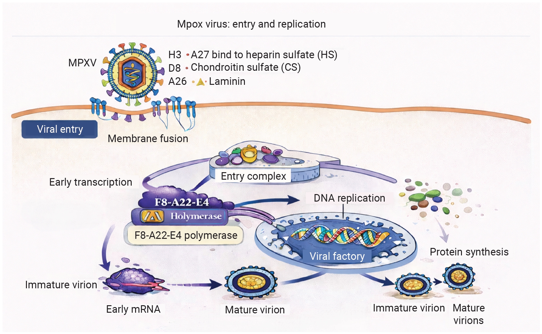
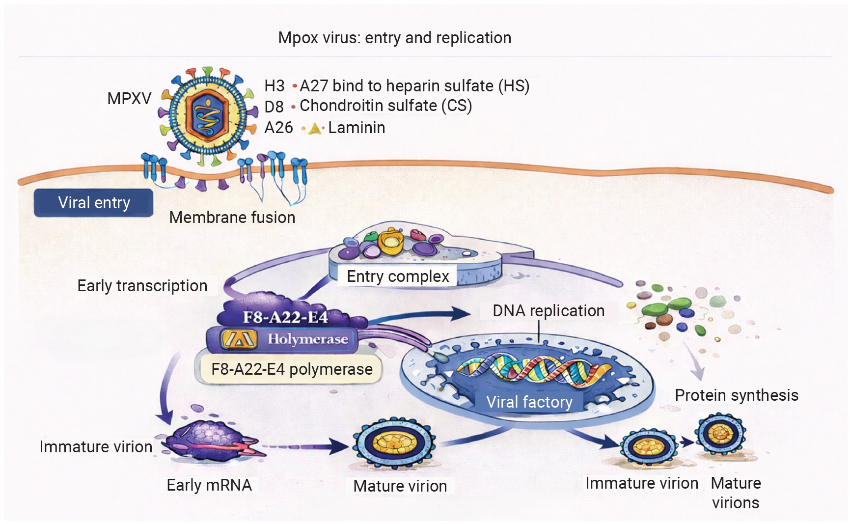
Viral entry is thought to occur through macropinocytosis, endocytosis, or cell membrane fusion. Several proteins are associated with viral attachment, including H3 and A27, which bind to heparan sulfate; D8, which binds to chondroitin sulfate; and A26, which binds to laminin. H3 is conserved among poxviruses. Another protein, A17, helps anchor A27, which interacts with A26 [
30]. Up to 12 proteins have been implicated in formation of the viral entry complex [
30,
31].
After infection, viral replication occurs in the cytoplasm and involves the Golgi apparatus. Cell fusion may also occur, leading to syncytium formation. Viral replication is mediated by the F8-A22-E4 polymerase. This holoenzyme is also involved in proofreading, thereby contributing to replication fidelity (
Figure 2). However, specific original research examining the viral entry and replication of atypical mpox remains scarce at the time of writing.
After local replication of mpox virus at the inoculation site, the virus spreads to the lymph nodes, leading to primary viremia during an incubation period lasting 1–3 weeks [
12,
32]. In an aerosol transmission model using cynomolgus monkeys, lymphatic spread through monocyte-associated viremia was demonstrated. Necrotic lesions and intracytoplasmic inclusions were also observed. The study further showed increased viral dissemination associated with secondary bacterial infection [
33]. After the initial spread, clinical symptoms begin with the prodrome, which corresponds to secondary viremia.
Clinical Manifestations
Prodromal Symptoms
The incubation period of mpox ranges from 5 to 24 days, with an average of 12 days [
14,
22]. In classical mpox infection, common systemic features often precede the development of a generalized rash. In atypical mpox, prodromal symptoms have been reported in 36% to 74.8% of patients [
6,
13,
14]. Prodromal symptoms may be completely absent, as described in case reports of patients with itchy vesicular genital lesions who had no prodromal symptoms and were initially misdiagnosed with herpes [
7,
24,
34]. When present, prodromal symptoms include fever, which occurs in 59%–62% of cases and may have sudden onset; lethargy, reported in 41%; myalgia, reported in 31%; sore throat, reported in 22%; and headache, reported in 27%. Lymphadenopathy has been observed in 50.6%–64.9% of cases and may be painful [
5,
7,
13,
20–
22,
24,
35–
41]. The prodrome may also be prolonged in some individuals [
6]. Case reports have also described mild diarrhea, pruritus, throat pain, generalized aches including chest pain, asthenia, and chills [
7,
36,
39–
43].
Lesions and Other Clinical Features
The characteristic rash of classical mpox usually begins as macular lesions and progresses through papular, vesicular, pustular, and crusting stages [
13,
36]. In atypical mpox, this typical evolution may not occur, and macular rashes may also be present [
7,
22]. Patients may present at any stage of lesion development, with asynchronous lesions or mixed crops [
14,
16,
22,
24,
26,
41,
44–
47]. In 2 large case series, skin lesions were present in 77%–95% of infected individuals [
4,
5]. The anogenital area was the most common site of these lesions, affecting 73% of patients with rashes [
4,
7,
22,
23,
25,
34,
47–
49]. Anogenital rashes were distributed as follows: genital lesions, 53%–56.4%; perianal lesions, 34%–41.6%; and genital and/or perianal lesions, 73%–88.3% [
13,
21]. Other commonly affected sites included the eyes, oropharynx, trunk, arms, or legs (55%); face (25%); and palms and soles (10%). In rare cases, paronychia has also been reported [
4,
13,
23,
24,
36,
50,
51].
A variety of lesion types have been observed, with vesiculopustular lesions being the most common (58%). Most patients reported fewer than 20–25 lesions (85%–93%) [
13]. Itching was associated with lesions in some cases [
7,
22]. Morphologically, pseudopustules, defined as whitish papules with a necrotic center, have been identified at the inoculation site in some cases. These are solid inflammatory lesions rather than true pustules containing purulent fluid [
13]. Public erythema, diarrhea, and odynophagia have also been reported [
7,
23]. Some individuals presented with a single genital ulcer or genital vesicular lesions, which could lead to misdiagnosis as another sexually transmitted infection [
4,
22,
35]. Verrucous lesions have also been reported [
25,
42].
Complications have been reported in approximately 13% of cases. These include severe pain at the site of anogenital lesions, proctitis, penile edema, phimosis or paraphimosis, secondary infections, pharyngitis, bronchopneumonia, encephalitis and other neurological complications, ocular complications including vision loss, acute kidney injury, abscess, sepsis, and myocarditis [
4,
19,
22,
48,
52–
55]. Differences from classical mpox, absence of prodromal symptoms, and clinical overlap with sexually transmitted infections make diagnosis and treatment of atypical mpox challenging. Patients often present to dermatology clinics, sexually transmitted infection clinics, and emergency departments [
4,
22]. These clinical settings are potential targets for enhancing early diagnosis and management of atypical mpox.
Diagnostic Approaches
Because atypical mpox differs clinically from classical disease, clinicians should recognize its key differential diagnoses. Patients presenting with vesicular lesions have been misdiagnosed with herpes or varicella-zoster virus infection and treated accordingly, often without substantial improvement [
7,
35,
56]. Single genital lesions have also been reported in several cases and may be misdiagnosed as syphilis [
4]. Case reports have described umbilicated lesions mimicking molluscum contagiosum [
36], impetigo, and erythema multiforme [
52]. The rash may also be confused with drug reactions, which can present as polymorphic eruptions. Therefore, a detailed history and a high degree of clinical suspicion in susceptible patients may reduce unnecessary laboratory investigations and facilitate diagnosis of atypical mpox.
The definitive diagnostic modality for atypical mpox is polymerase chain reaction (PCR). Because PCR is highly specific, clinicians must maintain a high degree of suspicion to diagnose atypical cases reliably. Samples for PCR can be obtained from crusts, lesion fluid, or swabs of cutaneous and anogenital lesions, which have a positivity rate of 97%. Other sample types include nasopharyngeal specimens, urine, semen, and blood, with positivity rates of 26%, 3%, 90%, and 7%, respectively. Thus, mucocutaneous lesion specimens are the most reliable samples, followed by those from seminal fluid [
4,
5,
7,
21,
35,
39,
57].
Dermoscopy has identified lesions with varying degrees of vascularity [
58]. Morphologically, lesions have shown a hemorrhagic center surrounded by a whitish ring, which is further surrounded by erythema. Pustular and vesicular lesions have also been observed. Histologically, Guarnieri inclusion bodies are seen in the cytoplasm. Lymphocytic infiltrates are present in lesions, along with full-thickness epidermal necrosis and ballooning degeneration of keratinocytes [
13,
59].
Serological tests, such as enzyme-linked immunosorbent assay and fluorescent antibody testing, can also be performed. Immunoglobulin M can be detected from day 5 to day 56 after rash onset, whereas immunoglobulin G can be identified from the end of the first week of illness [
52]. Recently, a method for rapid identification of antibodies against A29L, a surface antigen of mpox virus, was developed using high-throughput B-cell sequencing [
60]. However, these tests are not suitable for screening because they are typically performed only after rash onset. A transient decrease in T cells and atypical lymphocytes has also been reported [
61–
63]. In addition, leukocytosis, thrombocytopenia, decreased serum albumin levels, elevated transaminase levels, and increased blood urea nitrogen levels may be observed [
39]. Viral isolation and cell culture can be performed for research purposes in specialized high-biosafety-level laboratories [
21,
39,
64], but they have a limited role in routine diagnosis. Microbial cell-free DNA (mcfDNA) sequencing has also been explored for mpox diagnosis. This approach relies on peripheral blood examination and therefore can be performed before active lesions appear. Sequencing using mcfDNA can also accurately identify coinfections with other viruses and bacteria [
65].
Management Strategies
Treatment
Because clinical presentations vary, patients may require extensive supportive and symptomatic management. Isolation and home-based treatment are recommended for patients with mild symptoms [
4]. Pain control remains the mainstay of management for proctitis and lesion-associated pain [
5,
29,
66]. One report described a patient who required patient-controlled intravenous analgesia, whereas gabapentin and opioids have been used for short-term management of severe pain [
22,
29]. Topical lidocaine and stool softeners have also been used to manage proctitis [
66]. Oropharyngeal lesions may be managed with salt-water rinses, chlorhexidine mouthwash, or local analgesic mouthwash [
29,
66]. Topical and systemic antimicrobials, such as mupirocin, may be useful for mucocutaneous lesions with secondary bacterial infection, and corticosteroids may be used to manage local swelling around genital lesions [
17,
35,
66]. Antibiotics have been used for secondary bacterial infections [
5,
17,
29,
66]. Pruritic rashes have been managed with antihistamines [
5]. Several cases, accounting for 27%–29% of patients, have also involved concomitant sexually transmitted infections, which should be managed according to their respective protocols and treatment strategies [
4,
5,
18,
19,
26,
27,
67,
68]. Coinfection with cytomegalovirus has also been reported [
69].
Beyond symptomatic management, no treatment options are specifically approved for human mpox infection [
29]. Antivirals such as tecovirimat, cidofovir, and the cidofovir prodrug brincidofovir have been considered for treatment of human mpox and have been used in some cases [
16,
49,
70]. However, no clinical studies have confirmed the efficacy and safety of these antivirals for human mpox. Several new drugs are being investigated preclinically for mpox management [
70]. Tecovirimat, which is approved by the US Food and Drug Administration (FDA) for smallpox, is under investigation for treatment of mpox [
29,
70]. In the United States, it is available only through the Study of Tecovirimat for Human Monkeypox Virus (STOMP) trial [
29,
71]. The study is being conducted in the United States, Brazil, Peru, and Thailand. Tecovirimat has also been used successfully in some case reports [
4,
70]. Brincidofovir is another antiviral approved for use against smallpox and has shown promising results in preclinical studies [
29,
70]. In the United States, brincidofovir is available to clinicians only through FDA authorization for single-case emergency investigational new drug use. It may be authorized for adult and pediatric patients, including neonates, who have severe disease or are at high risk for severe disease or recrudescence [
29]. Cidofovir is another antiviral under consideration for mpox management. It is approved for treatment of cytomegalovirus retinitis in patients with acquired immunodeficiency syndrome. No studies have demonstrated the efficacy of cidofovir for mpox, and its use is further limited by severe renal toxicity [
29]. Vaccinia immune globulin intravenous (VIGIV) is also FDA-approved for management of vaccinia-related complications during immunization. Although no data are available on the safety and efficacy of VIGIV in human mpox infection, it has been reserved under the CDC expanded access protocol for management of orthopoxvirus outbreaks [
70]. It is also available on a case-by-case basis with CDC approval [
29,
70]. Trifluridine has been used with some success as adjunctive topical therapy in patients with ocular manifestations [
70].
Vaccination
Although no mpox-specific vaccine was initially available, the emergence of atypical mpox prompted efforts to deploy existing smallpox vaccines, which provide some cross-protection against mpox virus because of similarities between orthopoxviruses. Two orthopoxvirus vaccines, JYNNEOS (Bavarian Nordic A/S) and ACAM2000 (Emergent BioSolutions Inc.), are discussed by the CDC for use under specific conditions during human mpox outbreaks [
29,
70]. Both vaccines are live vaccines. JYNNEOS is based on a live attenuated vaccinia strain, modified vaccinia Ankara, whereas ACAM2000 contains replication-competent live vaccinia virus. JYNNEOS has been approved in various countries for mpox and smallpox. Two doses are recommended 28 days apart, with extension up to 35 days when necessary. In the United States, JYNNEOS has been approved for individuals at high risk for mpox. These include individuals with a new diagnosis of 1 or more sexually transmitted infections within the previous 6 months, more than 1 sexual partner, a history of sex at a commercial sex venue, or sexual intercourse associated with a large public gathering in an area where mpox transmission is occurring. Their sexual partners and people anticipating these exposures are also eligible for vaccination. Post-exposure prophylaxis is recommended within 4 days of exposure but may provide some benefit up to 14 days after exposure. Repeated doses are not recommended except for occupational exposure to orthopoxviruses; in such cases, booster doses can be given every 2–10 years. JYNNEOS is considered safer for immunocompromised individuals and pregnant patients because it does not replicate efficiently in humans. In contrast, ACAM2000 received emergency approval in the United States for the 2022 outbreak. ACAM2000 has more adverse effects and contraindications than JYNNEOS and carries a risk of vaccinia replication-related adverse events [
29,
70]. The MVA-BN and LC16M8 vaccines have been prequalified by WHO for use against mpox in Africa [
72,
73].
Challenges and Future Directions
The identification and management of atypical mpox face several major challenges, including divergent clinical presentations, stigma affecting populations at risk, limited clinician awareness, and the absence of mpox-specific treatment options [
74]. Atypical presentations, such as single lesions, asynchronous lesions, absent prodromal symptoms, and atypical rashes, complicate prompt recognition and diagnosis [
14,
16,
22,
24,
41,
44,
45]. Diagnosis is further hampered by co-occurring sexually transmitted infections. Healthcare providers in non-endemic regions often have limited familiarity with atypical mpox, which may delay diagnosis and treatment. Physicians in endemic areas generally have a higher degree of suspicion for mpox than those in non-endemic areas. Stigma and discrimination affecting certain communities, particularly MSM, may discourage individuals from seeking medical care until complications arise and may hinder contact tracing [
74]. In addition, limited access to diagnostic testing and the lack of mpox-specific antivirals may hamper case management.
Because large randomized controlled trials are lacking, several knowledge gaps remain regarding atypical mpox. Understanding the roles of asymptomatic and presymptomatic transmission and environmental contamination is necessary for establishing protocols to prevent mpox transmission comparable to that observed during the 2022 outbreak. Primary prevention measures for mpox infection also need to be better established; this requires clearer understanding of the pathophysiological mechanisms underlying transmission through various routes, particularly sexual contact. Although seminal fluid has tested positive in several studies, lesion swabs and lesion fluid also have high viral loads. Therefore, it is important to determine whether barrier protection during sexual contact would be effective in patients with anogenital lesions. Continuous genomic surveillance remains limited but is necessary to monitor viral evolution and its implications for transmissibility, virulence, and vaccine effectiveness. The long-term health consequences of atypical mpox are not yet fully understood, particularly in high-risk populations, because of the recency of the outbreak. As with coronavirus disease 2019, potential long-term complications and residual morbidities may occur and should be investigated in longitudinal studies.
The global spread of atypical mpox emphasizes the need for effective risk communication, efforts to combat stigma and discrimination, equitable access to healthcare, and strengthened surveillance systems. Collaboration among public health authorities, healthcare providers, researchers, and communities is necessary to address these challenges. International organizations and collaborations also play a crucial role in addressing knowledge gaps and ensuring effective global coordination.
This review is limited by its narrative design, reliance on predominantly observational studies and case reports, heterogeneity among published reports, and the rapidly evolving nature of the mpox literature, which may limit the generalizability and durability of some conclusions.
Future research priorities include ongoing genomic surveillance to monitor viral evolution, large-scale epidemiological studies to characterize transmission dynamics and zoonotic sources, and investigations of the long-term effects and potential complications of infection. Continued development of novel antiviral agents, immunomodulatory therapies, and next-generation vaccines is essential. Studies should also explore combination therapies for synergistic effects, assess asymptomatic transmission and environmental contamination, and evaluate long-term vaccine effectiveness and the need for booster doses. Measuring the effects of public health interventions and community engagement on outbreak control will help strengthen preparedness for future mpox outbreaks and other emerging infectious diseases. Through sustained research and comprehensive public health strategies, the global community can better address the challenges posed by atypical mpox.
HIGHLIGHTS
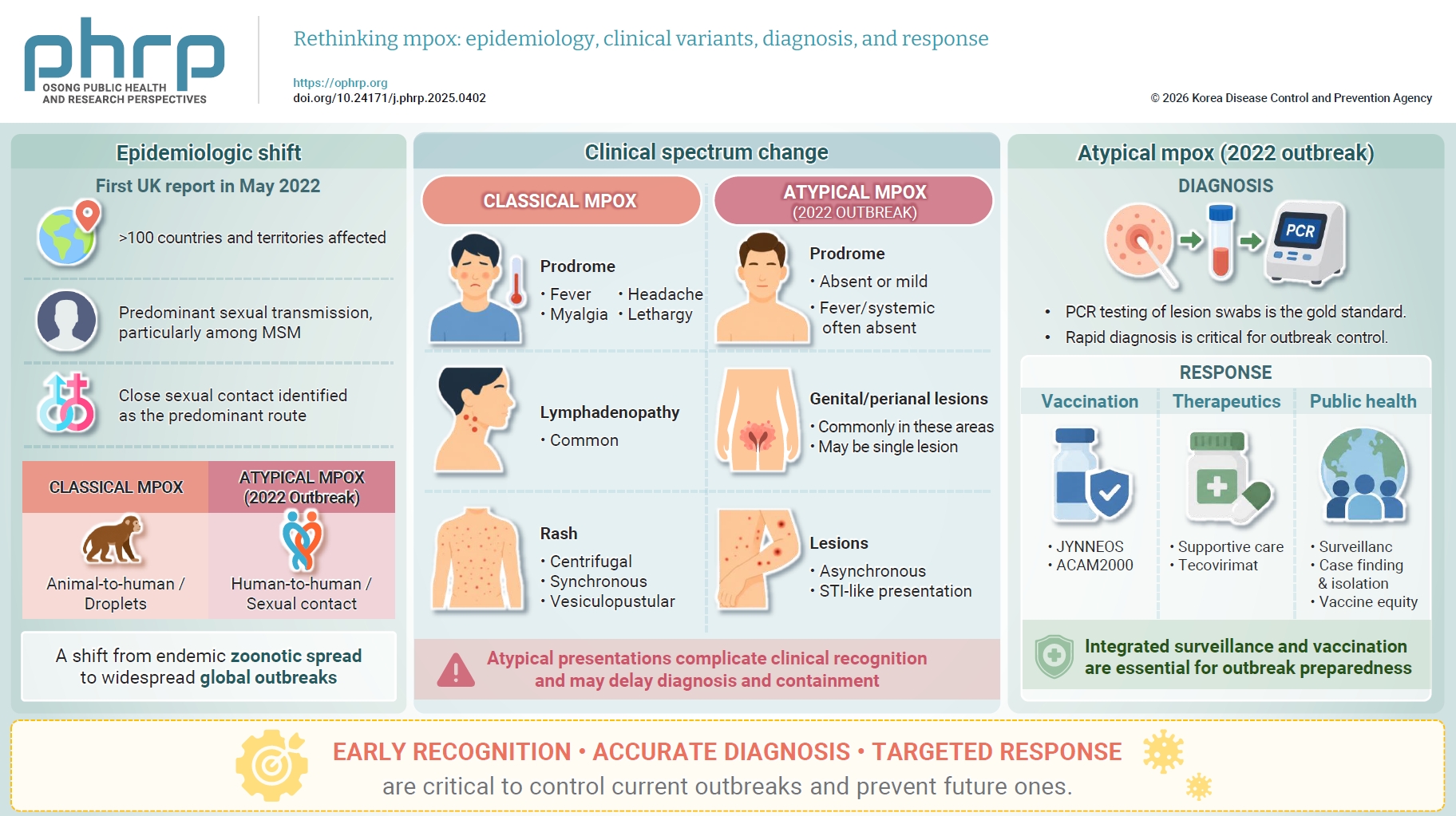
• The 2022 multinational mpox outbreak marked a shift in transmission dynamics, with sexual contact emerging as the predominant route of transmission.
• Atypical mpox often presents with genital and perianal lesions, absent or minimal prodromal symptoms, and asynchronous rash evolution.
• Molecular diagnosis, primarily polymerase chain reaction testing of lesion swabs, remains the gold standard for confirmation.
• Management focuses on supportive care and symptomatic relief, with antivirals such as tecovirimat available under specific protocols.
• Continued genomic surveillance and equitable vaccine distribution are essential for future outbreak preparedness.