Skip to contents Skip to Global Navigation Menu
  • KDCA
  • Contact us
  • E-Submission

PHRP : Osong Public Health and Research Perspectives

OPEN ACCESS. pISSN: 2210-9099. eISSN: 2233-6052
Original Article

Environmental emergence and dissemination of clinically relevant multidrug-resistant bacteria and resistance genes in sewage and aquatic ecosystems


Published online: July 23, 2026

1Department of Biosciences, Veer Narmad South Gujarat University, Surat, India

2Shree Ramkrishna Institute of Computer Education and Applied Sciences, Sarvajanik University, Surat, India

Corresponding author: Pravin Dudhagara Department of Biosciences, Veer Narmad South Gujarat University, Surat-395007, Gujarat, India E-mail: dudhagarapr@gmail.com
• Received: February 19, 2026   • Revised: May 11, 2026   • Accepted: May 28, 2026

© 2026 Korea Disease Control and Prevention Agency.

This is an open access article under the CC BY-NC-ND license (http://creativecommons.org/licenses/by-nc-nd/4.0/).

  • 1,030 Views
  • 53 Download
  • Objectives
    This study characterized multidrug-resistant (MDR) bacteria in sewage, river, and marine ecosystems in South Gujarat, India.
  • Methods
    Water samples were collected from 25 hospital drainage, sewage treatment/pumping, Tapi River, and coastal marine sites at multiple time points. From 270 screened colonies, 166 morphologically and biochemically distinct nonduplicate isolates were retained. Antimicrobial susceptibility was assessed, and isolates were classified as MDR, extensively drug-resistant (XDR), or pan-drug-resistant (PDR). Biofilm formation, metabolic activity, extracellular polymeric substance protein, heavy metal tolerance, extended-spectrum β-lactamase production, carbapenemase production, and metallo-β-lactamase activity were assessed phenotypically. Tapi River estuary water was used for taxonomic profiling and antibiotic resistance gene (ARG) detection.
  • Results
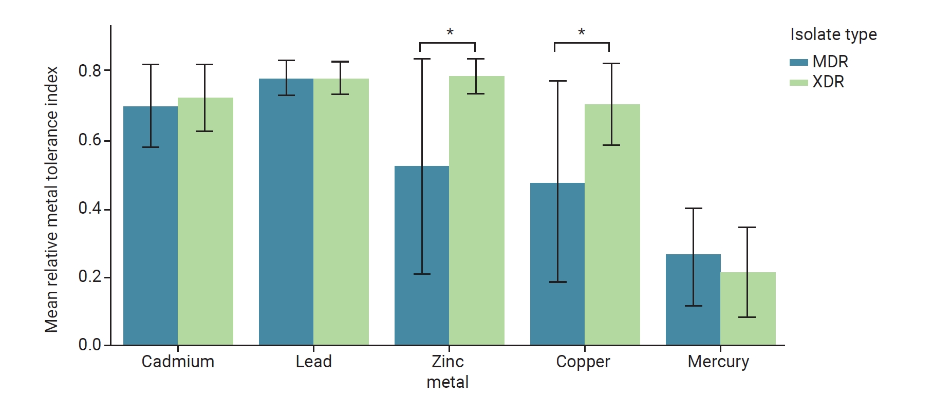
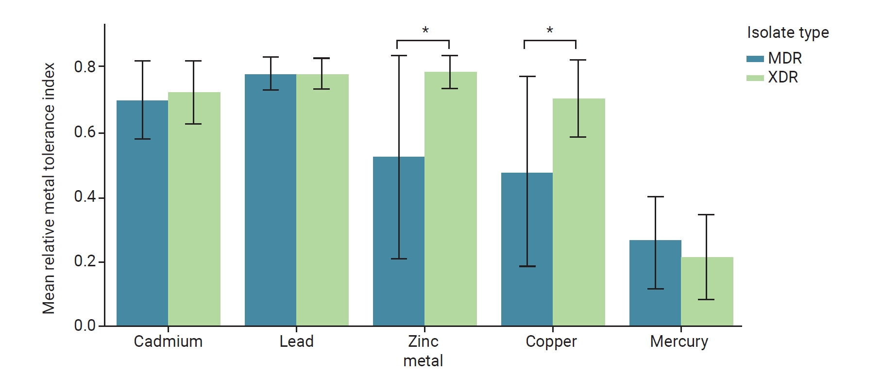
    The 166 isolates comprised 45 bacterial species, with clinically significant Gram-negative pathogens predominating, including Pseudomonas aeruginosa (n=24, 14.5%), Ochrobactrum intermedium (n=16, 9.6%), Stenotrophomonas maltophilia (n=15, 9.0%), and Escherichia coli (n=12, 7.2%). MDR phenotypes were detected in 80.1% of isolates; 18.1% were XDR, and 1.8% were PDR, with PDR isolates confined to river water samples. Biofilm formation was observed in 86.1% (n=143) of isolates, including 28.3% (n=47) strong, 24.7% (n=41) moderate, and 32.5% (n=54) weak producers. Extended-spectrum β-lactamase production was confirmed in 13.4% of Gram-negative isolates, and carbapenemase activity was detected in 11 isolates. Zinc and copper tolerance were significantly higher in XDR than in MDR isolates (p<0.05). Metagenomics identified efflux pumps as the dominant ARG class (36.2%), followed by target-site mutations (21.3%) and β-lactamases (14.9%), and detected blaCTX-M-15, blaTEM-207, gyrA, and parC.
  • Conclusion
    These interconnected aquatic systems represent important reservoirs for community-level antimicrobial resistance transmission.
Antimicrobial resistance (AMR) ranks among the leading causes of death worldwide [1]. Without international action, AMR-related deaths are expected to reach 10 million per year by 2050 [2]. Inappropriate antibiotic use in various settings contributes to AMR and facilitates the intersectoral transfer of resistant bacteria through food chains, wastewater systems, and natural ecosystems, creating interconnected resistance pathways that pose a One Health threat [3–5].
The World Health Organization (WHO) has identified AMR as an urgent One Health concern [6]. However, the extent to which clinical discharge points seed environmental reservoirs remains unclear. Hospital effluents are enriched with pathogenic and antibiotic-resistant organisms, potentially presenting a pathway for clinically controlled pathogens to enter municipal sewage systems [7,8].
Wastewater treatment plants may also harbor persistent resistant bacteria and antibiotic resistance genes (ARGs) that re-enter receiving ecosystems [9,10]. The increasing detection of extended-spectrum β-lactamase (ESBL)- and metallo-β-lactamase (MBL)-producing bacteria in hospital wastewater and surrounding aquatic environments highlights the environmental and public health implications of last-resort antibiotic resistance [11]. Sewage systems may function as resistance “melting pots,” receiving high loads of antibiotics, biocides, and metals that create selective pressures favoring the emergence of multidrug-resistant (MDR) and extensively drug-resistant (XDR) strains.
Riverine systems can act as reservoirs and conduits that transport MDR bacteria and ARGs while providing nutrient-rich environments that support bacterial survival, biofilm formation, and horizontal gene transfer [12]. Biofilms further enhance bacterial survival and persistence in aquatic environments. Biofilms are organized microbial communities within an extracellular polymeric substance (EPS) matrix, which protects cells from antimicrobial exposure and environmental stress while facilitating horizontal gene transfer [13,14]. Rivers in India are often contaminated with subinhibitory concentrations of antibiotics, promoting biofilm formation [15,16]. Similarly, marine and coastal waters receiving river discharge serve as ecological “memory banks” of AMR, enabling MDR pathogens to persist, recombine, and re-enter human and animal populations [17,18].
Environmental antibiotics, heavy metals, feces, and microplastics can promote co-selection, stress adaptation, and long-term AMR [19]. Heavy metals, including lead, cadmium, mercury, copper, and zinc, are persistent environmental pollutants common in industrial and municipal wastewater. Long-term exposure can impose sustained selective pressure on microbial communities and co-select for antibiotic resistance through shared resistance mechanisms [20,21].
Understanding environmental resistome reservoirs is important for infection prevention, antibiotic stewardship, and public health preparedness. Clinicians often encounter MDR infections in patients without conventional healthcare-associated risk factors, suggesting environmental acquisition [22]. The presence of priority pathogens such as Pseudomonas aeruginosa and ESBL-producing Escherichia coli in water bodies may create direct transmission pathways to humans [23,24]. Knowledge of local resistance patterns, such as the prevalence of blaCTX-M-15 in environmental reservoirs, is essential for optimizing empiric treatment regimens for community-acquired infections [25].
Among the most clinically dangerous bacterial threats are so-called “superbugs,” organisms that combine virulence with resistance to multiple antibiotic classes and often render standard treatment ineffective. The ESKAPEE pathogens—Enterococcus faecium, Staphylococcus aureus, Klebsiella pneumoniae, Acinetobacter baumannii, P. aeruginosa, Enterobacter spp., and E. coli—account for a disproportionate burden of hospital-acquired and community-acquired infections [26]. These organisms exhibit extraordinary genomic plasticity, including large, flexible accessory pangenomes that harbor mobile genetic elements, which enable rapid horizontal acquisition and transfer of resistance determinants [27,28]. This genetic fluidity allows ESKAPEE pathogens to assemble new resistance combinations under selective pressure, making them well adapted to antibiotic-rich environments, including hospital effluents and wastewater ecosystems [27].
Carbapenemase-encoding genes confer resistance to last-resort carbapenems for Gram-negative infections. Carbapenemases are classified into 3 Ambler molecular classes: class A serine carbapenemases (e.g., K. pneumoniae carbapenemase); class B metallo-beta-lactamases (e.g., New Delhi metallo-β-lactamase, verona integron-encoded metallo-β-lactamase, and imipenemase metallo-β-lactamase), which require zinc ions for activity; and class D oxacillinases (e.g., OXA-48 and OXA-23), which are serine-based but structurally distinct from class A enzymes [29,30]. Carbapenemase genes are increasingly detected in wastewater, river sediments, and coastal waters, where mobile elements may facilitate rapid interspecies spread [30,31]. Understanding the environmental genomic pool is therefore central to predicting and intercepting resistance at the human–animal–environment interface.
Sewage, river, and marine ecosystems at this interface are increasingly recognized as potential reservoirs for AMR emergence, persistence, and dissemination, but they remain insufficiently studied. Integrated phenotypic evidence linking AMR, biofilm formation, EPS production, and metal tolerance among cultivable bacteria in interconnected aquatic ecosystems remains limited. Consequently, this study analyzed bacteria recovered across a connected environmental gradient in South Gujarat, India, extending from hospital drainage systems and municipal sewage to the Tapi River and coastal discharge points. By combining AMR profiling with analyses of biofilm-related traits and heavy metal tolerance within a One Health framework, this study provides background evidence on environmental AMR hotspots and potential transmission routes relevant to human health.
Study Area and Sample Collection
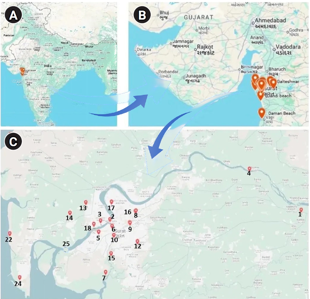
This study used no direct patient-level clinical samples. The study was conducted from January 2023 to June 2025 in and around Surat, South Gujarat, India. Sampling sites included hospital drainage systems representing clinical reservoirs; sewage treatment plants (STPs); sewage pumping stations (SPSs) within Surat city; upstream and downstream sites along the Tapi River; and marine environments, including estuarine and coastal sites. Samples were collected aseptically in sterile containers, transported at 4 °C, and processed within 24–48 hours. Sewage samples were collected multiple times per month and pooled to generate composite samples. Sampling locations are shown in Figure 1. For metagenomic analysis, multiple water samples from the surface to approximately 3 feet were collected in sterile 1-L bottles from the Tapi River estuary (site 25; downstream of all STPs/SPSs; Figure 1C), in June 2025 to capture the cumulative resistome load entering the marine ecosystem.
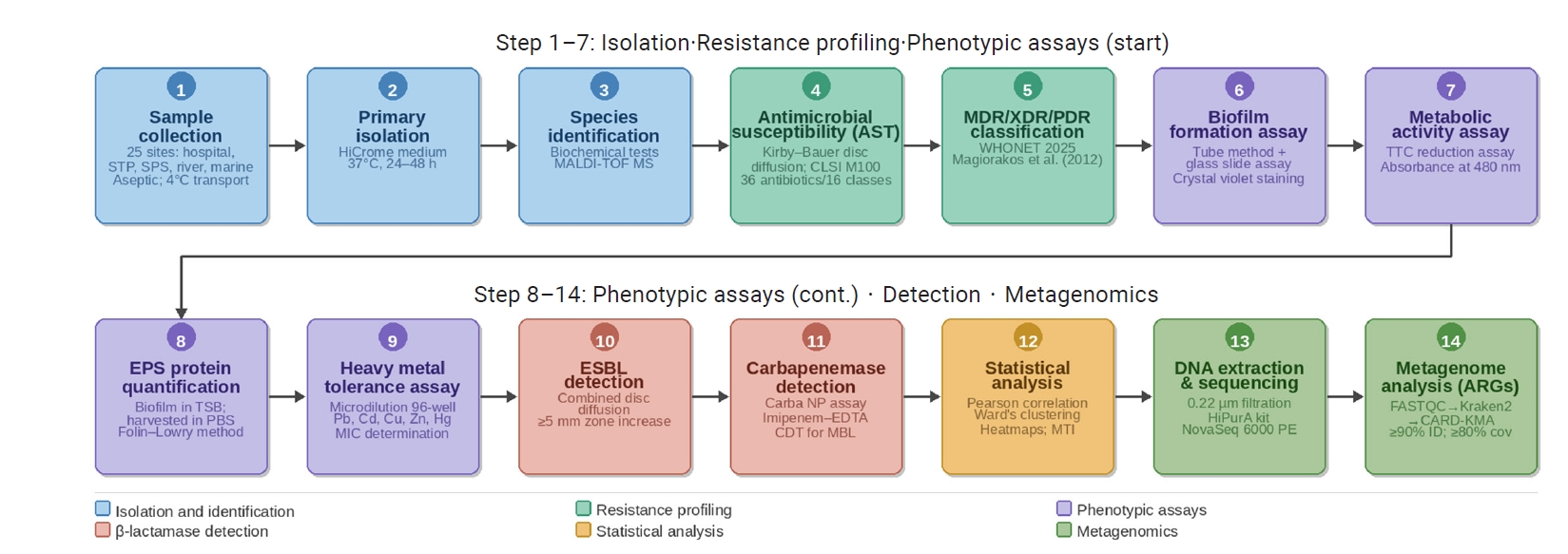
The methodological workflow (Figure 2) began with sample collection and matrix-assisted laser desorption/ionization time-of-flight mass spectrometry (MALDI-TOF MS) identification. Isolates then underwent antimicrobial susceptibility testing (AST) for MDR classification, along with phenotypic assays for biofilm formation, metabolic activity, and heavy metal tolerance. Specific resistance mechanisms were characterized before statistical analysis and metagenomic sequencing on the NovaSeq 6000 platform. Finally, bioinformatic analysis using the Comprehensive Antibiotic Resistance Database-K-mer Alignment (CARD-KMA) pipeline identified ARGs.
Isolation and Identification of Bacterial Isolates
A loopful of each collected sample was streaked onto HiCrome Universal Differential Medium (HiMedia Laboratories Pvt. Ltd.; catalog No. M1600) and incubated aerobically at 37 °C for 24–48 hours. Morphologically distinct colonies were selected and subcultured onto nutrient agar for purification by successive single-colony streaking. Purified isolates underwent preliminary identification using standard biochemical tests, including Gram staining, catalase, oxidase, motility, indole, methyl red-Voges-Proskauer, citrate utilization, triple sugar iron, urease activity, and gelatin hydrolysis assays, performed according to MacFaddin [32]. Final species-level identification was performed by MALDI-TOF MS. A pure culture from a single colony was processed according to the manufacturer’s guidelines for the autoflex maX system. Briefly, a colony was mixed with matrix solution, such as α-cyano-4-hydroxycinnamic acid, spotted onto the MALDI plate, and air-dried. The plate was inserted into the MALDI-TOF MS instrument, the sample was laser-ionized, the time of flight was measured, and the resulting spectra were matched against the database. Identification was completed within minutes [33].
AST and Resistance Profiling
Antimicrobial susceptibility was determined using the Kirby–Bauer disc diffusion method on Mueller-Hinton agar (MHA) [34–36]. (Bacterial suspensions adjusted to a 0.5 McFarland standard were swabbed evenly onto MHA plates. Antibiotic discs (HiMedia Laboratories Pvt. Ltd.) from multiple classes were placed at defined intervals, and the plates were incubated at 37 °C for 18–24 hours. To assess the relevance to human health, results were interpreted strictly according to Clinical and Laboratory Standards Institute (CLSI) M100 clinical breakpoints [37–39]. The antibiotics, disc potencies, and classes used in this study are detailed in Table 1.
Susceptibility data were analyzed using WHONET 2025 software to classify isolates as MDR, XDR, or pan-drug-resistant (PDR) according to the standardized international expert definitions of Magiorakos et al. [40]. MDR was defined as acquired non-susceptibility to at least 1 agent in 3 or more antimicrobial categories, XDR was defined as non-susceptibility to at least 1 agent in all but 2 or fewer antimicrobial categories, and PDR was defined as non-susceptibility to all agents in all antimicrobial categories [40].
Biofilm Formation and Metabolic Activity
Biofilm production was assessed qualitatively by inoculating 5 mL of tryptic soy broth (TSB) with 10 µL of test culture, followed by incubation at 37 °C for 48 hours. After decanting, the tubes were washed with phosphate-buffered saline (PBS; pH 7.2) and deionized water and then air-dried. The tubes were stained with 0.1% crystal violet for 15 minutes and inverted to dry. Biofilm formation was confirmed by the presence of a visible layer on the tube walls, whereas a simple ring at the air–liquid interface was interpreted as a negative result. Biofilm formation was scored as negative (0), weak (1), moderate (1+), or strong (2+) [41].
Metabolic activity was assessed directly in the biofilms developed by each strain using colorimetry [42]. Reduction of 2,3,5-triphenyltetrazolium chloride (TTC) was measured spectrophotometrically at 480 nm. A 10-µL aliquot of test culture was inoculated into 5 mL of TSB and incubated at 37 °C for 48 hours. After incubation, the biofilm was scraped and resuspended in 1 mL of PBS. After 50 µL of TTC dye was added and the suspension was incubated for 3 hours at room temperature, 100 µL of the solution was transferred to a 96-well plate. Optical density (OD) was measured at 480 nm.
EPS Protein Estimation
EPS extraction was performed using the method described by Jiao et al. [43]. Bacteria were cultured in TSB at a 1:100 dilution and incubated at 37 °C for 3 days. After incubation, the supernatant was decanted, and the tubes were washed 3 times with PBS (pH 7.2). The surface-attached biofilm was scraped into 1 mL of PBS using a sterile scalpel and gently vortexed to generate a dispersed EPS solution [43]. Protein content in the EPS solution was quantified using the Folin-Lowry method, with bovine serum albumin as the standard [44,45]. Briefly, 1 mL of EPS solution was mixed with 5 mL of alkaline copper solution and incubated for 30 minutes at room temperature. After 0.5 mL of Folin-Ciocalteu reagent was added and the mixture was incubated for an additional 15 minutes, absorbance was measured at 750 nm using a spectrophotometer.
Heavy Metal Tolerance Assay
Tolerance to lead, cadmium, copper, zinc, and mercury was evaluated using a broth microdilution assay in 96-well plates, following the approaches described by Pagnucco et al. [46] and Ojo et al. [47]. Briefly, metal chloride salts were dissolved in sterile distilled water and filter-sterilized through 0.22-µm membranes to prepare stock solutions. Serial 2-fold dilutions were prepared in nutrient broth at 12.5, 25, 50, 100, 200, 400, 600, and 800 ppm for lead, cadmium, copper, and zinc and 3.12, 6.25, 12.5, 25, 50, 100, and 200 ppm for mercury, reflecting the higher toxicity of mercury at lower concentrations. Bacterial metabolic activity in each well was assessed using TTC dye, and OD was measured at 480 nm after incubation at 37 °C for 24 hours. The minimum inhibitory concentration was defined as the lowest metal concentration that completely inhibited TTC reduction, indicating the absence of detectable metabolic activity.
Detection of ESBL and Carbapenemase Production
Gram-negative isolates resistant to 1 or more third-generation cephalosporins (cefotaxime, ceftazidime, or ceftriaxone) were screened for ESBL production using the combined disc diffusion method [48]. Discs containing cefotaxime (30 µg) and ceftazidime (30 µg) were placed alongside their respective clavulanic acid combination discs (cefotaxime/clavulanic acid, 30/10 µg; ceftazidime/clavulanic acid, 30/10 µg) on MHA plates inoculated with a 0.5 McFarland bacterial suspension. Plates were incubated at 37 °C for 18 hours, and inhibition zone diameters were measured. An increase of ≥5 mm in the inhibition zone diameter around a clavulanic acid–containing disc compared with the corresponding cephalosporin disc alone was interpreted as confirmatory for ESBL production, in accordance with CLSI guidelines. CLSI-recommended reference strains of E. coli, P. aeruginosa, S. aureus, and K. pneumoniae maintained in the institutional culture collection were used as quality control organisms for AST, ESBL detection, and carbapenemase assays according to CLSI M100 guidelines [37–39]. Appropriate sterility and uninoculated media controls were included in all phenotypic assays.
Gram-negative isolates with reduced susceptibility or resistance to 1 or more carbapenem antibiotics (imipenem, meropenem, or ertapenem) were investigated for carbapenemase production. The Carba NP assay described by Nordmann et al. [48] was used as the primary confirmatory test for carbapenemase activity. Solution A (0.05% phenol red and 0.1 mmol/L ZnSO₄•7H₂O, pH 7.8) and solution B (6 mg/mL imipenem/cilastatin in solution A) were prepared. A concentrated bacterial inoculum was prepared in 100 µL of sterile water and divided into 2 tubes, labeled A and B. Solution A (100 µL) was added to tube A, and solution B (100 µL) was added to tube B; the tubes were then incubated at room temperature for 2 hours. A color change from red to yellow in tube B, reflecting imipenem hydrolysis and a subsequent pH decrease, indicated a positive result [49]. Carbapenem-resistant isolates were screened for MBL production using the imipenem–ethylenediaminetetraacetic acid (EDTA) combined disc test. Bacterial suspensions adjusted to 0.5 McFarland were swabbed onto MHA. Two 10-µg imipenem discs were placed 20 mm apart, and 10 µL of 0.5 M EDTA was added to one disc. After overnight incubation, an increase of ≥7 mm in the inhibition zone around the imipenem-plus-EDTA disc compared with the imipenem disc alone was considered positive for MBL production [49].
Statistical Analysis
Statistical analyses, including Pearson correlation analyses with significance set at p<0.05, were performed using Microsoft Excel 2019 (Microsoft Corp.). Hierarchical clustering with Ward linkage and Euclidean distance, along with heatmaps, was used for visualization. Bacterial tolerance to cadmium, lead, zinc, copper, and mercury was assessed by normalizing growth against the lowest concentration. A concentration-weighted index was calculated to quantify stress adaptation:
Metal tolerance index=(∑(Ci×Gi))/(∑Ci×100),
where Ci represents the metal concentration and Gi denotes the relative growth (%). This approach generated a normalized score from 0 to 1, and composite indices were derived by averaging individual metal scores.
DNA Extraction, Library Construction, and Shotgun Metagenomic Sequencing
A total of 100 mL of river water was pooled and filtered through a presterilized 0.22-µm filter to capture microorganisms. Biomass retained on the filter was used for DNA isolation with the HiPurA Water DNA Purification Kit (MB577; HiMedia Laboratories Pvt. Ltd.). DNA quality was assessed by 0.8% agarose gel electrophoresis, and DNA was quantified using a Qubit 4 Fluorometer (Thermo Fisher Scientific). Shotgun metagenomic libraries were constructed using the QIAseq FX DNA Library Kit (Qiagen), and sequencing was performed using paired-end chemistry (250×2) on an Illumina NovaSeq 6000 platform (Illumina) at the Gujarat Biotechnology Research Centre shared next-generation sequencing facility, Gujarat, India.
Analysis of Metagenome Sequences for Decoding the River Microbiome and ARGs
FASTQ files generated by sequencing were processed using FastQC (v0.11.9) for quality assessment [50]. Taxonomic classification of quality-filtered reads was performed using Kraken2 (v2.1.2) [51] within the Galaxy platform against the standard Kraken2 database. Classification results were visualized as interactive multilayered pie charts of microbial taxa using Krona (v2.8) [52]. For ARG identification, quality-controlled metagenomic reads were mapped against the Comprehensive Antibiotic Resistance Database (CARD; v3.2.7) [53] using the K-mer Alignment (KMA) algorithm (v1.4.9) [54] integrated within the Bacterial and Viral Bioinformatics Resource Center (BV-BRC) framework [55]. ARG–species associations were inferred using default BV-BRC/KMA alignment parameters, with sequence identity >70% considered for ARG assignment.
Isolation, Characterization, and Identification of Bacteria
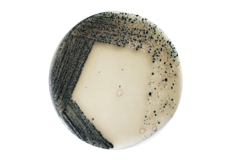
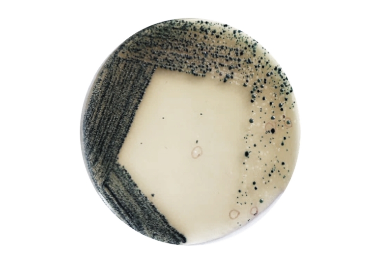
A total of 270 colonies were screened on HiCrome Universal Differential Medium, and 166 morphologically and biochemically distinct nonduplicate isolates were retained for detailed characterization (Figure 3). Gram staining indicated a predominance of clinically relevant Gram-negative pathogens over Gram-positive isolates. From this initial collection, a subset of 166 isolates was selected for comprehensive phenotypic characterization (Table S1). Selection criteria included morphologically unique colonies that varied in color, size, shape, elevation, and texture. Isolates were selected from all sampling sites. The 166 isolates were subcultured onto nutrient agar and purified through successive single-colony streaking. These purified strains underwent preliminary identification using standard biochemical tests, with final species-level confirmation by MALDI-TOF MS (Table S2).
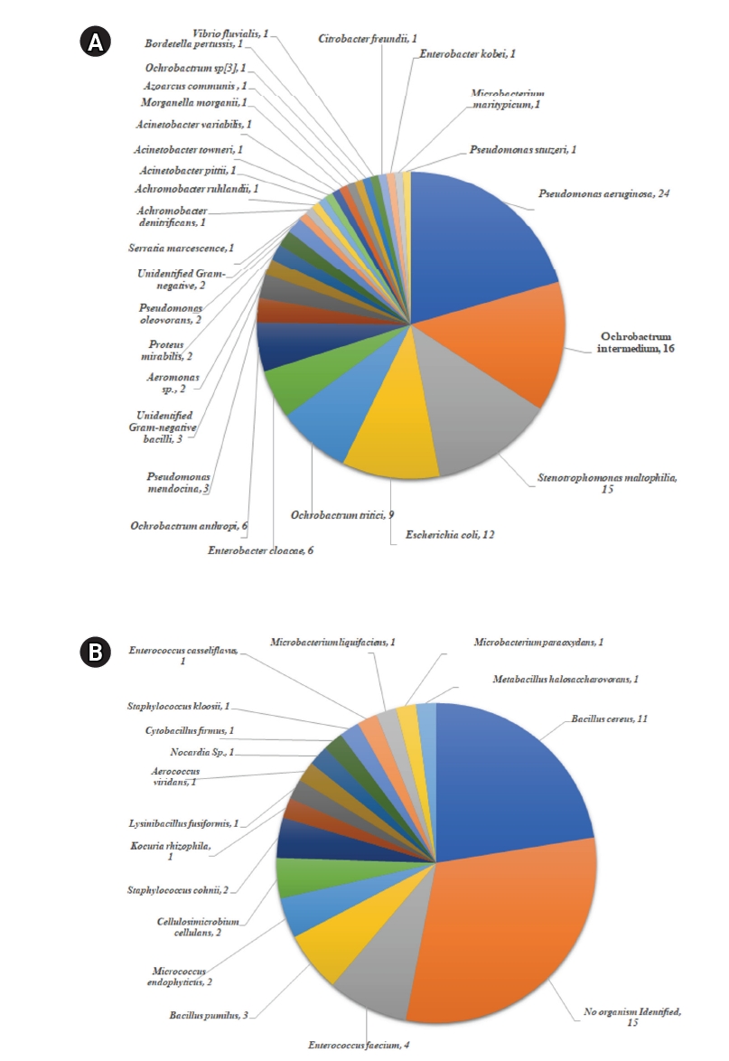
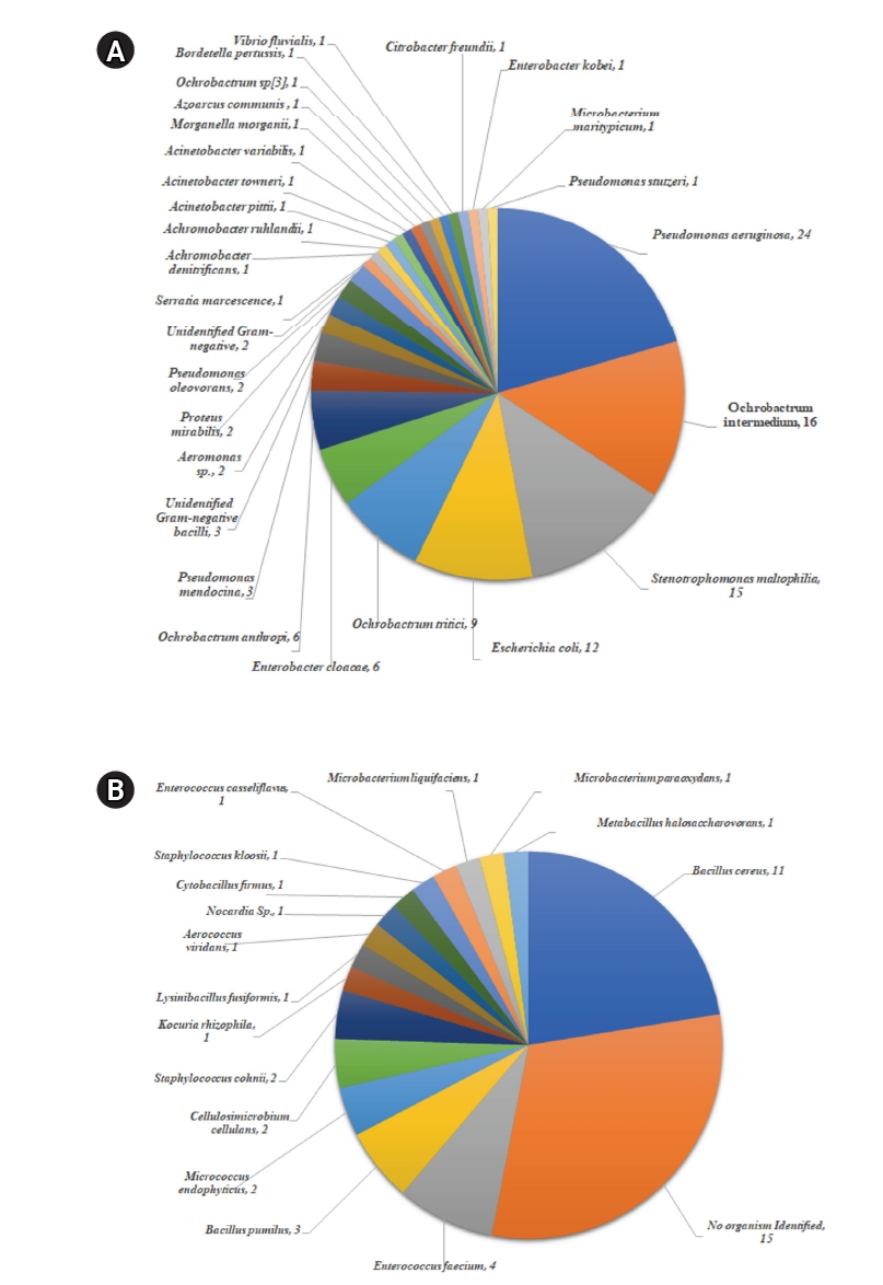
MALDI-TOF MS identified 45 distinct bacterial species (Figure 4). Gram-negative isolates were dominated by P. aeruginosa (n=24), Ochrobactrum intermedium (n=16), Stenotrophomonas maltophilia (n=15), and E. coli (n=12). Additional Gram-negative species included Ochrobactrum tritici (n=9), Ochrobactrum anthropi (n=6), and Enterobacter cloacae (n=6), while other genera, such as Acinetobacter and Achromobacter, occurred at lower frequencies. Among Gram-positive isolates, Bacillus cereus (n=11) was the dominant species, followed by E. faecium (n=4) and Bacillus pumilus (n=3). Rare isolates of Micrococcus and Staphylococcus were observed at low frequencies, and 15 isolates could not be identified to the species level.
Antibiotic Susceptibility Analysis using WHONET
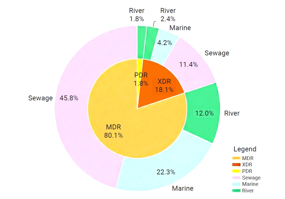
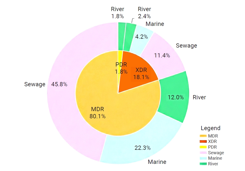
A total of 166 bacterial isolates were tested against multiple antibiotic classes, and susceptibility data (Table S3) were analyzed using WHONET software (Table S4). Based on standardized definitions, isolates were classified as MDR, XDR, or PDR (Figure 5). MDR isolates predominated (80.1%) and were distributed across all environmental sources; sewage accounted for the largest proportion (45.8%), followed by marine (22.3%) and river (12.0%) samples. XDR isolates comprised 18.1% of the population and were primarily associated with sewage (11.4%), with smaller contributions from marine (4.2%) and river (2.4%) environments. PDR isolates represented 1.8% of the total and were detected only in river water samples. These PDR isolates exhibited resistance profiles that would severely compromise all standard therapeutic options.
Phenotypic Profiling and Metabolic Correlation of Biofilm-Forming MDR Isolates
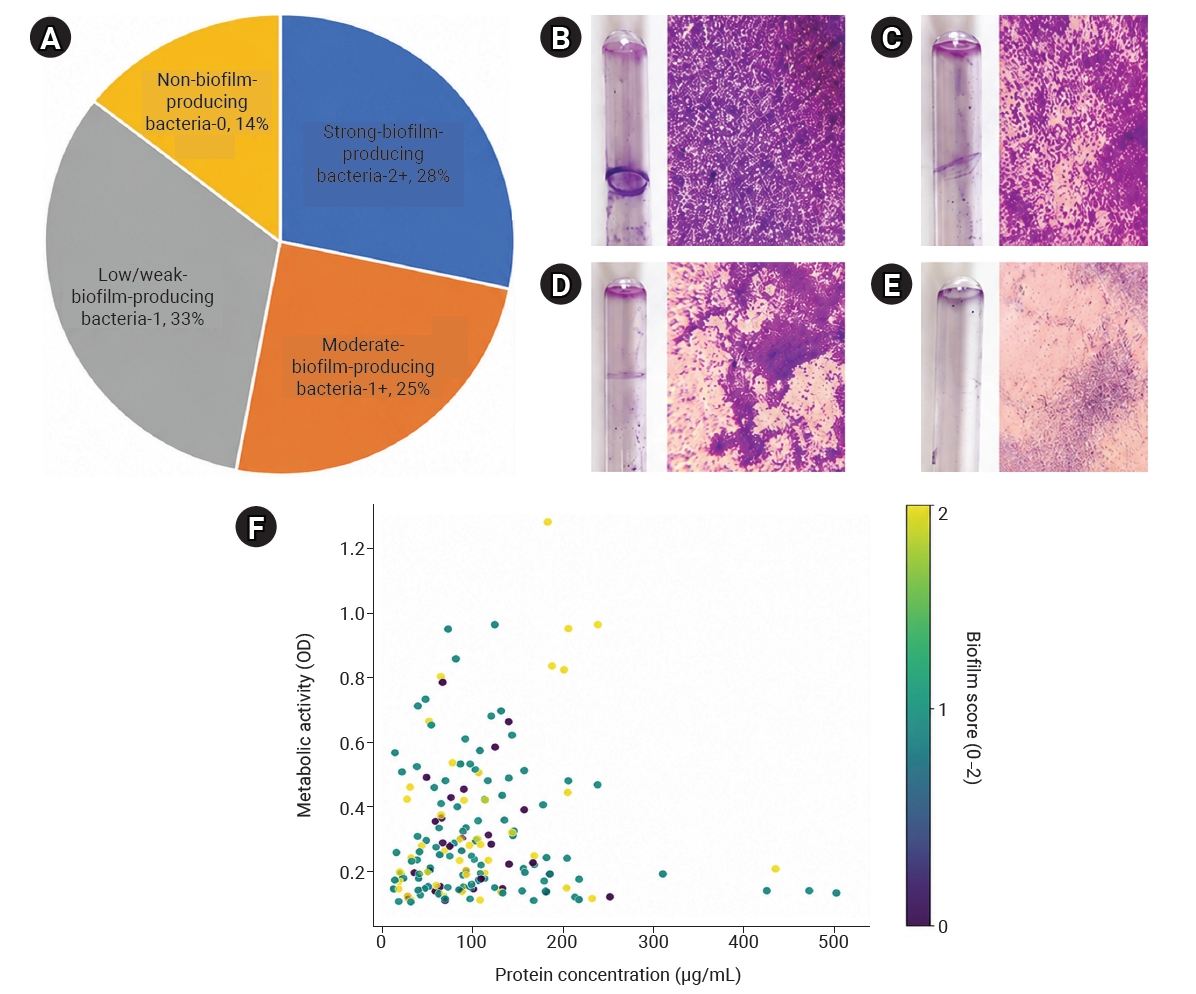
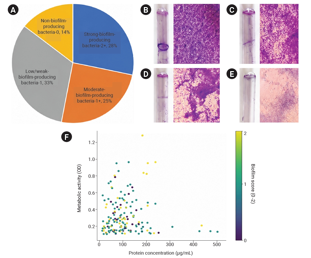
Biofilm-forming potential was assessed in 166 MDR isolates using qualitative tube and glass slide methods. Most isolates exhibited some degree of biofilm formation (Figure 6A). Specifically, 28% (n=47) were classified as strong biofilm producers, 25% (n=41) as moderate producers, and 33% (n=54) as low or weak producers, and 14% (n=24) were non-biofilm producers.
Macroscopically, strong biofilm producers exhibited a thick, continuous crystal violet–stained ring at the air–liquid interface, whereas weak producers showed only faint staining. Microscopy (×100 magnification) corroborated these observations, revealing dense, compact cellular aggregates in strong producers but sparse or absent surface-associated cells in weak and non–biofilm-forming isolates (Figure 6B–E).
To characterize physiological heterogeneity, the relationship between metabolic activity, measured as OD at 480 nm, and bacterial biomass, measured as protein concentration, was examined. The data were widely dispersed and lacked linearity (Figure 6F). No significant relationship was found between metabolic activity and biomass accumulation (Pearson correlation r=−0.002, p=0.98; Spearman rank correlation ρ=0.02, p=0.75). However, isolates with higher metabolic activity (OD at 480 nm >0.8) were predominantly strong biofilm producers but had moderate biomass levels (<250 µg/mL), whereas isolates with the highest biomass (>450 µg/mL) consistently showed low metabolic activity (OD at 480 nm <0.2).
Correlation Analysis of Metal Tolerance Indices and Physiological Attributes in Isolates
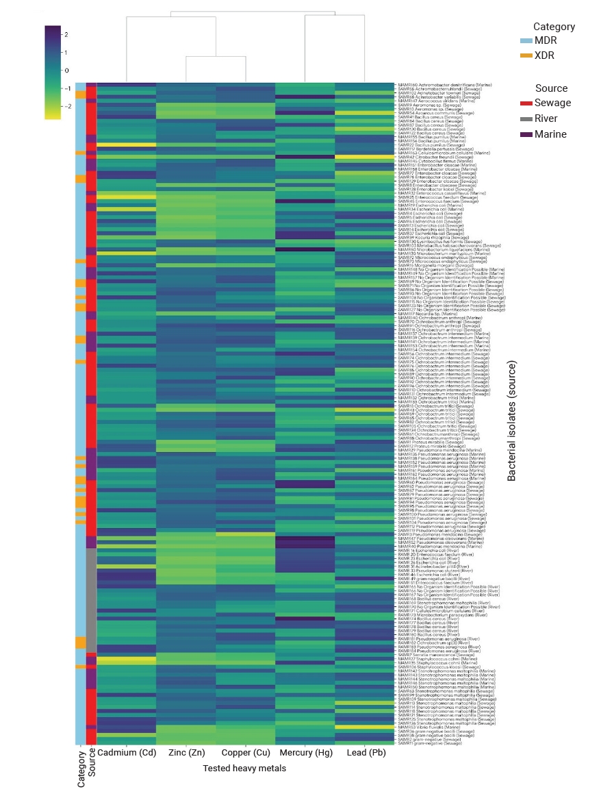
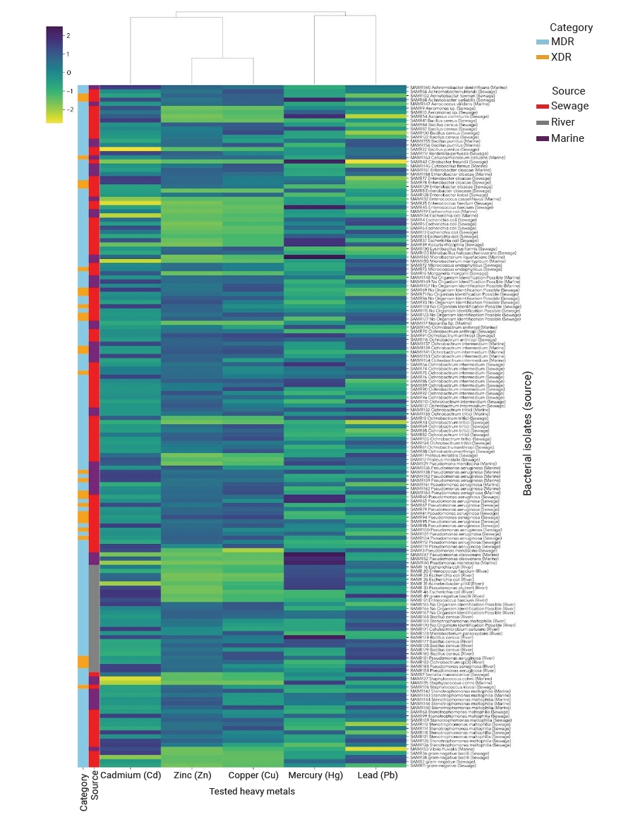
Comparative analysis of MDR, XDR, and PDR isolates from marine, river, and sewage environments revealed distinct patterns by environmental source and resistance category (Figure 7). Zinc and lead tolerance consistently exceeded cadmium and mercury tolerance, whereas copper tolerance varied by source (Figure 8). Marine and sewage isolates exhibited higher and more stable composite metal tolerance indices (CMTIs) than river isolates, and XDR and PDR groups showed greater tolerance stability than MDR groups.
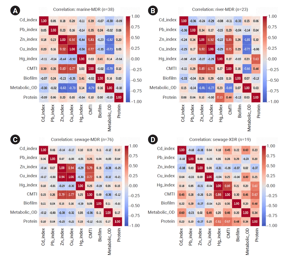
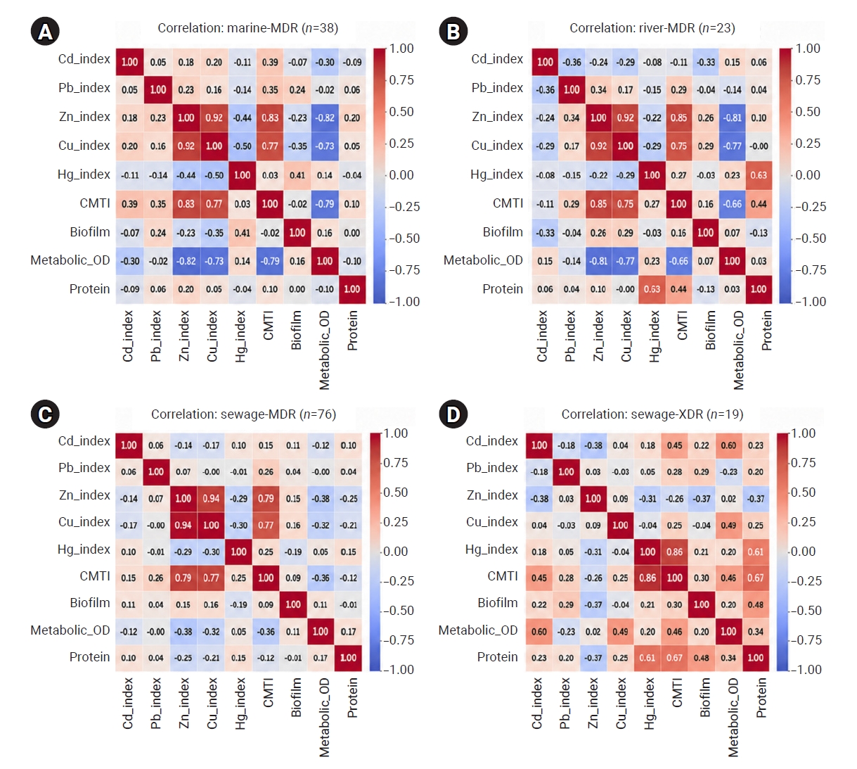
Zinc and copper showed strong positive correlations with MDR groups (r=0.92–0.94) and with CMTI across marine (r=0.77–0.83), river (r=0.75–0.85), and sewage (r=0.77–0.79) isolates. In contrast, cadmium showed inconsistent associations (r=−0.11 to 0.39). Mercury emerged as a major contributor only in sewage XDR isolates, showing strong correlations with CMTI (r=0.86) and protein content (r=0.61). Biofilm formation was primarily source-dependent, with sewage isolates consistently scoring higher. Although biofilm formation was decoupled from metal tolerance in MDR populations (|r|=0.24), sewage XDR isolates showed positive correlations between biofilm formation and both CMTI (r=0.30) and protein content (r=0.48). XDR and PDR isolates showed lower metabolic activity than MDR isolates across all environmental sources. Metabolic activity was negatively correlated with metal tolerance in MDR isolates from marine (r=−0.73 to −0.82), river (r=−0.66 to −0.81), and sewage (r=−0.32 to −0.38) environments (Figure 9). However, this trade-off was attenuated in sewage XDR isolates, in which metabolism was positively associated with cadmium (r=0.60) and copper (r=0.49).
Prevalence of ESBL and Carbapenemase-Producing Isolates
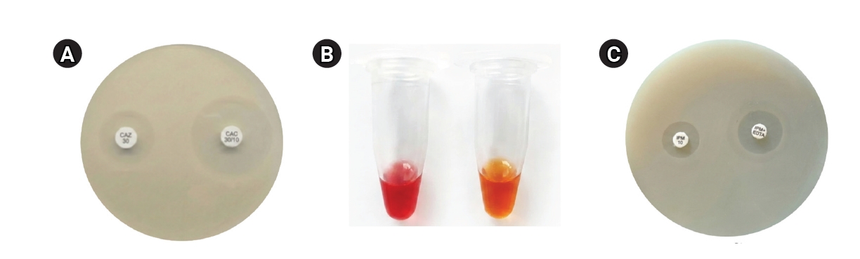
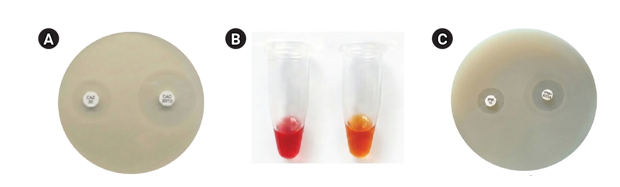
Among the 166 characterized bacterial isolates, 142 Gram-negative strains were screened for β-lactamase-mediated resistance using phenotypic assays. ESBL production was detected in 19 isolates (13.38%) by the combined disc diffusion method, as indicated by enhanced inhibition zones around ceftazidime–clavulanic acid compared with ceftazidime alone (Figure 10A). ESBL prevalence was highest among sewage-derived isolates, followed by marine isolates, whereas no ESBL producers were detected in river water isolates. ESBL-producing isolates were taxonomically diverse and included Pseudomonas spp., E. coli, S. maltophilia, and members of the genera Citrobacter, Morganella, and Acinetobacter.
Carbapenem resistance was investigated in 84 Gram-negative isolates. Confirmatory testing using the Carba NP assay identified 11 isolates (13.1%) as confirmed carbapenemase producers (Figure 10B). To classify the specific resistance mechanism, the imipenem–EDTA combined disc test was used. Although the imipenem–EDTA combined disc test suggested potential MBL activity in 60 isolates (71.4%), only the 11 isolates confirmed by the Carba NP assay were classified as true carbapenemase producers (Figure 10C). This approach minimized the potential for phenotypic false-positive findings associated with older methods or non-enzymatic resistance mechanisms.
Taxonomic Composition and Distribution of Microbiota in the River Metagenome
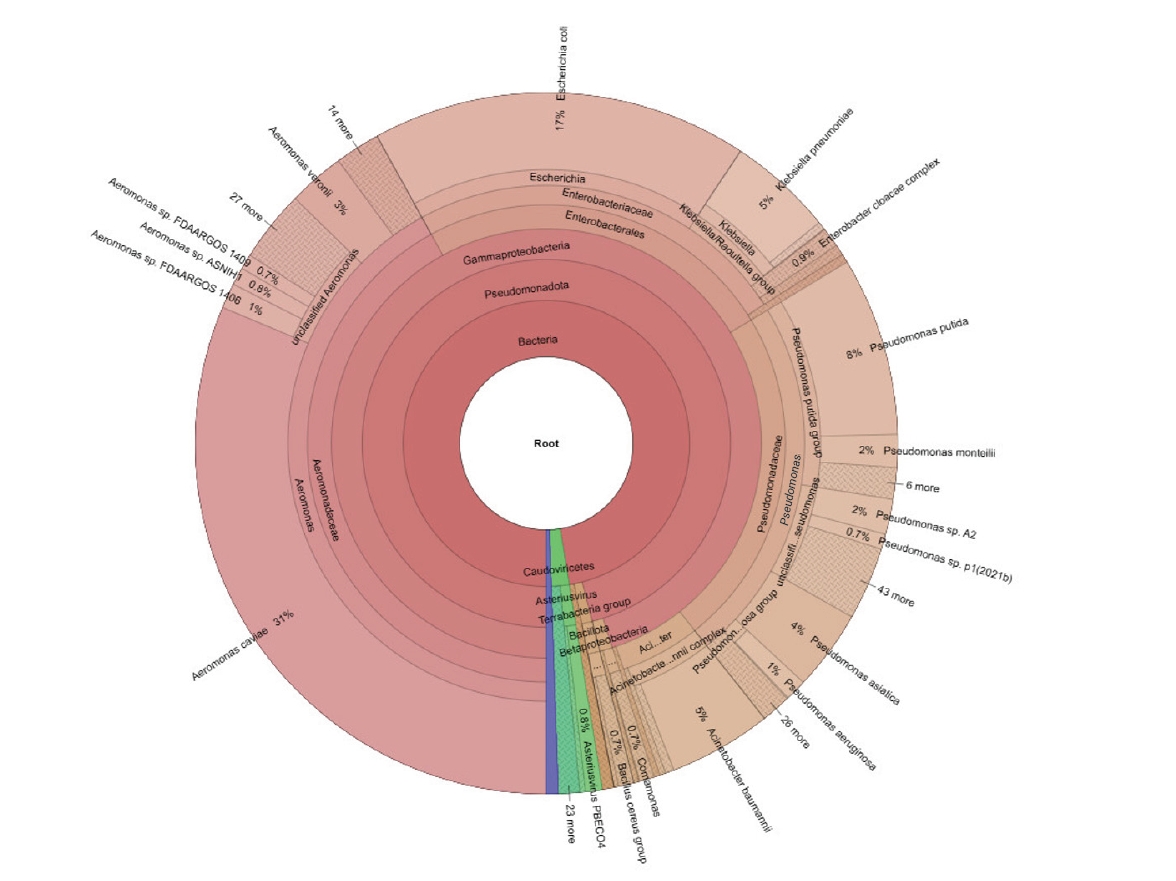
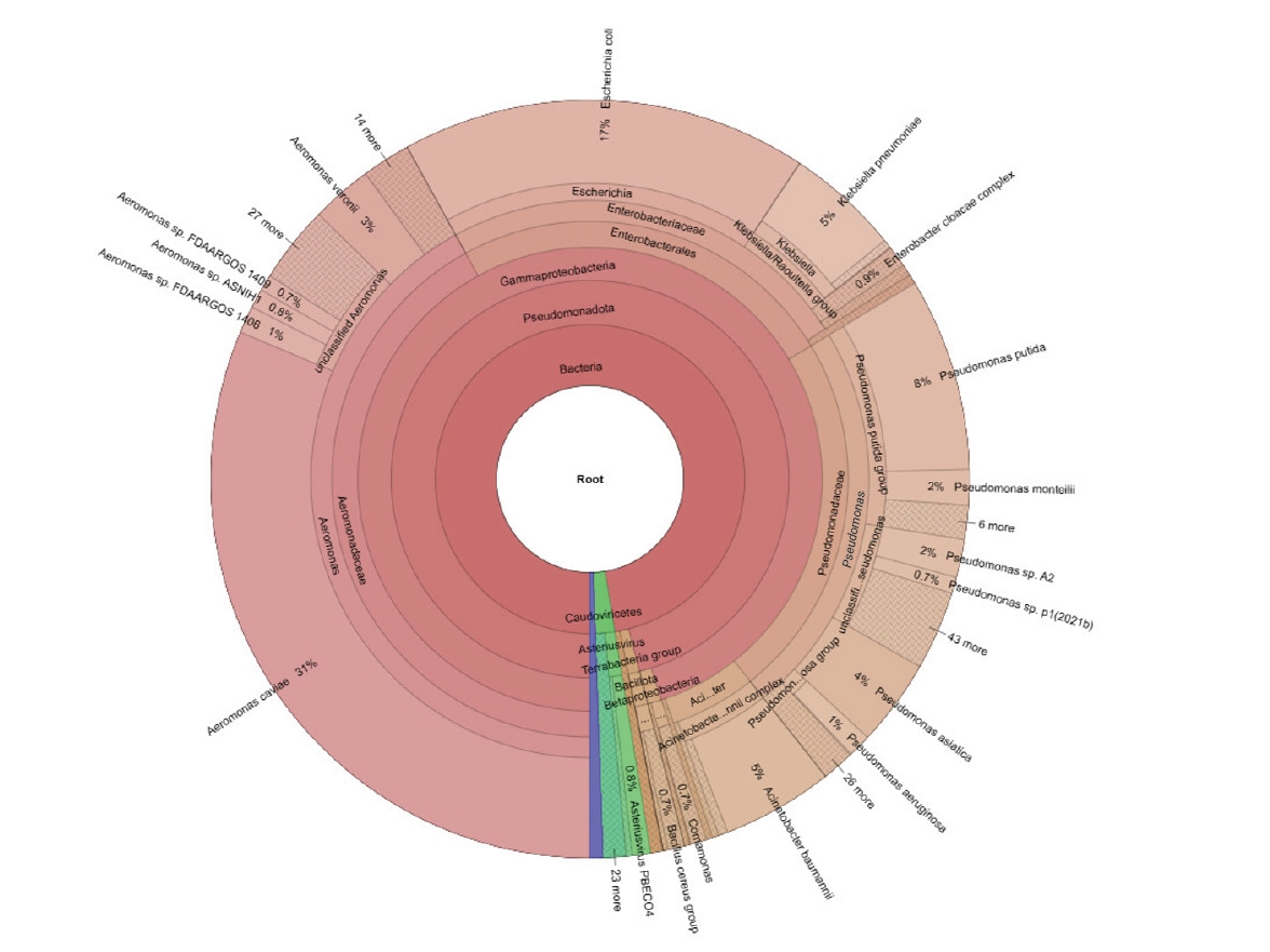
Taxonomic profiling revealed that the community was strongly dominated by Pseudomonadota, with minor contributions from Bacillota and other groups (Figure 11). At the species level, Aeromonas caviae was most abundant, representing 31% of the total population and reflecting the influence of the marine and riverine sampling environments.
Clinically relevant members of the family Enterobacteriaceae constituted a major fraction of the community and were led by E. coli (17%) and K. pneumoniae (5%). Their presence coincided with sampling locations influenced by treated and untreated wastewater discharge. The family Pseudomonadaceae was also prominent, represented by Pseudomonas putida (8%) and the opportunistic pathogen P. aeruginosa (5%). A. baumannii and E. cloacae complex were also identified at lower relative abundances. The remaining fraction of the community comprised diverse environmental genera, including Comamonas and unclassified Aeromonas species, underscoring the complex microbial ecology of these sites. The co-occurrence of autochthonous aquatic bacteria, such as Aeromonas, with typical fecal indicators and nosocomial pathogens, such as E. coli, K. pneumoniae, and P. aeruginosa, indicates that native aquatic bacteria and clinically relevant opportunistic pathogens coexist in these environments. Metagenomic analysis detected the globally dominant ESBL gene blaCTX-M-15 and fluoroquinolone resistance mutations in gyrA, confirming the presence of high-risk clinical resistance determinants in the river microbiome.
Detection of ARGs
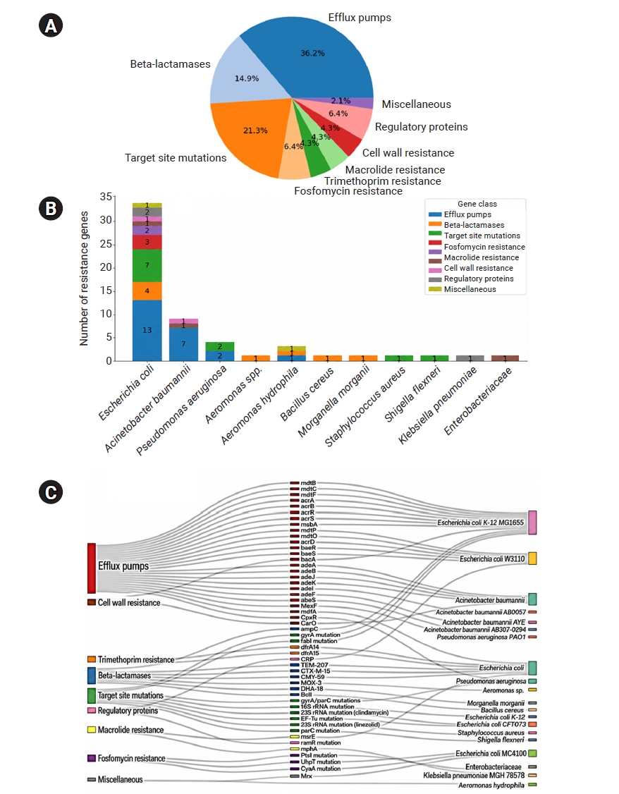
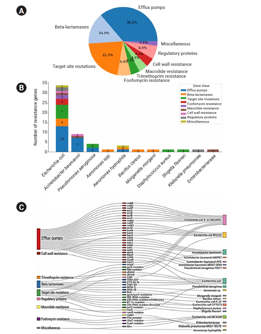
CARD analysis identified multiple ARG classes with more than 70% sequence identity. Efflux pump genes represented the largest ARG class (36.2%), followed by target-site mutations (21.3%) and β-lactamases (14.9%) (Figure 12A). Resistance determinants associated with efflux pumps were consistently detected across multiple taxonomic groups, whereas β-lactamase and target-site mutation genes showed more restricted taxonomic distributions. E. coli harbored the highest number and diversity of ARG classes; A. baumannii contained 2 ARG classes, while Aeromonas hydrophila and P. aeruginosa each contained 1 ARG class (Figure 12B).
The detected ARGs encompassed resistance mechanisms targeting multiple antibiotic classes, including β-lactams, fluoroquinolones, aminoglycosides, macrolides, and fosfomycin, reflecting broad-spectrum resistance potential within the environmental metagenome. Efflux pumps were the dominant resistance mechanism, with numerous genes, including acrA, acrB, members of the mdt and ade families, and MexF, distributed across E. coli, A. baumannii, P. aeruginosa, and A. hydrophila. Numerous β-lactamases, including TEM-207, CTX-M-15, CMY-59, MOX-3, DHA-18, BcII, and ampC, were detected in E. coli, Aeromonas sp., Morganella morganii, and B. cereus. Target-site mutations, including rrsB, gyrA/parC, fabI, and EF-Tu, were concentrated in E. coli, S. aureus, Shigella flexneri, and P. aeruginosa. Fosfomycin resistance genes (PtsI, UhpT, and CyaA) were restricted to E. coli MC4100, whereas trimethoprim resistance genes (dfrA14 and dfrA15) were found only in E. coli. Macrolide resistance genes (mphA and msrE) were observed in Enterobacteriaceae and E. coli, and cell wall resistance genes (bacA and CarO) were identified in E. coli and A. baumannii. Regulatory resistance determinants, including CRP and ramR mutations, were detected among E. coli and K. pneumoniae, whereas the miscellaneous resistance gene mrx was detected only in A. hydrophila. Among the detected taxa, E. coli harbored the highest diversity of resistance gene classes. In contrast, A. baumannii, A. hydrophila, and P. aeruginosa exhibited narrower distributions of resistance gene classes, dominated primarily by efflux-related and β-lactam-associated mechanisms (Figure 12C; Table S5).
This study demonstrated a high prevalence of MDR bacteria across all investigated aquatic environments, with sewage-derived isolates exhibiting the highest resistance burden. Comparable patterns have been reported globally, with wastewater systems receiving clinical, industrial, and agricultural inputs acting as convergence points for resistant bacteria [7,56]. The predominance of MDR phenotypes observed here is consistent with reports from untreated urban effluents in Europe [57].
The overlap in resistance patterns among environmental isolates, including WHO critical priority pathogens [23], indicates that resistance determinants are widely distributed across aquatic compartments. Similar observations have been documented in both developed and developing regions [58–60]. Sewage-associated bacteria exhibited higher and more consistent biofilm-forming capacity than river and marine isolates, likely reflecting sustained exposure to antibiotics, disinfectants, and chemical pollutants [61]. The persistence of biofilm-forming MDR isolates in sewage effluents suggests that conventional wastewater treatment processes may be insufficient to eliminate biofilm-associated pathogens, supporting tertiary treatment protocols that target aggregate-associated bacteria [62]. Marine isolates such as O. tritici and S. maltophilia displayed EPS levels comparable to those reported for strong clinical biofilm producers. Robust EPS production may represent an adaptive response to osmotic stress caused by salinity, which may also select for traits that enhance survival and colonization in the human host [63]. Thus, robust biofilm formation is not restricted to healthcare-associated bacteria [64].
Strong biofilm formation was not consistently associated with the highest AMR profiles. This is consistent with studies showing that environmental parameters can influence biofilm formation independently of resistance gene carriage [65,66].
Heavy metal tolerance profiling provided further evidence of antibiotic–metal co-selection. A substantial proportion of MDR isolates exhibited tolerance to elevated lead concentrations, with additional tolerance to copper and zinc observed in several isolates. The zinc/copper tolerance–biofilm correlation suggests a multilayered response to environmental stress. This co-occurrence may reflect the activity of promiscuous efflux pumps, such as P-type ATPases or resistance–nodulation–division family systems [67]. However, this study demonstrated only statistical associations, and the association between metal exposure and biofilm intensity may be driven by oxidative-stress responses. Heavy metals induce reactive oxygen species protection, upregulating cyclic di-GMP intracellular signaling pathways [68]. Elevated cyclic di-GMP levels act as a switch, repressing flagellar motility and stimulating the synthesis of EPS and curli fimbriae [69]. This results in a thick biofilm matrix that creates a physical barrier against antimicrobial agents and actively sequesters metal ions through electrostatic interactions [70].
Metagenomic detection of resistance determinants provides ecosystem-level context for these phenotypic observations. Efflux systems identified within the resistome contribute to cross-resistance against antibiotics and heavy metals, consistent with the observed correlations between zinc, copper, and CMTIs in cultured isolates [21]. The phenotypic detection of ESBL-producing isolates (13.4%) and carbapenemase-producing organisms confirmed by the Carba NP assay (11 isolates; 13.1% of carbapenem-resistant Gram-negative isolates tested) is notable alongside the metagenomic identification of β-lactamase gene classes, including blaCTX-M-15, blaCMY-59, blaDHA-18, and blaampC, in the river resistome. Carbapenemase confirmation in this study was performed exclusively using the Carba NP assay; the modified Hodge test was not used because it was deprecated by CLSI in 2018 because of its poor specificity and high false-positive rate [30]. The higher number of combined disc test–positive isolates (60 isolates, 71.4%) compared with Carba NP-confirmed producers (11 isolates) is consistent with published observations that the imipenem–EDTA disc test can yield false-positive results [30]. However, the presence of specific ARGs did not always correlate linearly with phenotypic resistance levels [71]. The presence of diverse β-lactamase genes in river water metagenomes suggests that these resistance determinants are embedded within broader environmental microbial communities.
From a One Health perspective, the combined phenotypic and metagenomic findings emphasize the interconnectedness of sewage, river, and marine ecosystems as shared AMR reservoirs. The presence of priority pathogens in riverine systems is particularly concerning for human populations [24]. The high prevalence of XDR phenotypes and blaCTX-M-15 genes in river water suggests that environmental exposure may contribute to rising rates of community-acquired ESBL infections [25]. Consequently, empiric therapy with third-generation cephalosporins for waterborne infections in this region may carry a high risk of treatment failure [72]. Aquatic environments may function as active reservoirs for MDR organisms. The detection of diverse ARGs in river water highlights the role of riverine ecosystems as dynamic incubators for resistance traits under fluctuating environmental conditions [73].
Limitations
Several limitations should be noted. First, this study did not use a systematic longitudinal sampling design. Second, the study was conducted in a single geographic region and included limited metagenomic sampling. Third, these findings were based on phenotypic and genomic associations, and experimental verification of the underlying molecular mechanisms was beyond the scope of this study. In addition, antibiotic or heavy metal concentrations were not quantified. Finally, this study did not prove direct transmission to individual patients.
This study showed that sewage, river, and marine ecosystems are interconnected reservoirs and potential evolutionary hotspots of MDR, XDR, and PDR bacteria, most notably Gram-negative opportunistic pathogens with AMR, biofilm-forming capacity, metabolic heterogeneity, and environmentally relevant heavy metal tolerance. The close association between zinc and copper tolerance and advanced resistance phenotypes is consistent with possible metal-associated co-selection, highlighting the potential importance of non-antibiotic stressors in shaping environmental AMR patterns. Detection of ESBL- and carbapenemase-producing isolates, together with a heterogeneous aquatic resistome characterized by efflux pumps, target-site mutations, and β-lactamase genes, supports the interpretation that aquatic environments are active sites of resistance persistence and potential dissemination. These findings clarify key ecological and functional drivers of AMR in interconnected aquatic environments and underscore the urgent need to incorporate environmental monitoring into AMR prevention efforts within a broader One Health framework.
• Multidrug-resistant (MDR) bacteria, including clinically relevant pathogens, are prevalent across interconnected sewage and aquatic ecosystems in South Gujarat.
• High rates of MDR and extensively drug-resistant phenotypes were identified among environmental isolates, with prevalence of 80.1% and 18.1%, respectively.
• Zinc and copper tolerance were significantly associated with extreme resistance profiles, suggesting possible metal-driven co-selection.
• Metagenomic profiling confirmed the presence of clinically important resistance genes, including blaCTX-M-15 and blaTEM 207, in the river microbiome.
• These aquatic systems may constitute important reservoirs for community-acquired infections, supporting the need for urgent environmental public health surveillance.
Supplementary data are available at https://doi.org/10.24171/j.phrp.2026.0186.
Table S1.
Isolate details across various sample source.
j-phrp-2026-0186-Supplementary-Table-S.xlsx
Table S2.
Matrix-assisted laser desorption/ionization time-of-flight score for the isolate’s identification.
j-phrp-2026-0186-Supplementary-Table-S.xlsx
Table S3.
Antibiotics sensitivity profile of the isolates
j-phrp-2026-0186-Supplementary-Table-S.xlsx
Table S4.
Antibiogram of isolates across drug classes
j-phrp-2026-0186-Supplementary-Table-S.xlsx
Table S5.
Metagenomic read mapping report using Comprehensive Antibiotic Resistance Database
j-phrp-2026-0186-Supplementary-Table-S.xlsx

Ethics Approval

Not applicable.

Conflicts of Interest

The authors have no conflicts of interest to declare.

Funding

This research was funded by the Gujarat State Biotechnology Mission (GSBTM) under the “Network Program on Antimicrobial Resistance, Superbugs, and One Health” project (No. GSBTM/JD(R&D)/616/21-22/1236) for the 2021–22 financial year. The authors acknowledge the invaluable support provided by GSBTM throughout this project.

Availability of Data

The datasets generated during and/or analyzed during this study are available in the supplementary file.

Authors’ Contributions

Conceptualization: SA, RP, PD; Data curation: SA, NV, AV, AG; Formal analysis: SA, KA, NV, AV; Funding acquisition: RP, PD; Investigation: SA, KA, NV, AV; Methodology: SA, KA, NV, AV; Project administration: RP, PD; Resources: RP, PD; Supervision: RP, PD; Visualization: SA, NV, AV, AG; Writing–original draft: SA; Writing–review & editing: all authors. All authors read and approved the final manuscript.

Acknowledgements

This study was conducted under the “Network Program on Antimicrobial Resistance, Superbugs, and One Health” project (No. GSBTM/JD(R&D)/616/21-22/1236), under the Research Support Scheme 2021–22 of the Gujarat State Biotechnology Mission, Gandhinagar, India. The authors sincerely thank the Bioinformatics and Supercomputer Lab at the Department of Biosciences, Veer Narmad South Gujarat University, for providing computational resources and technical assistance.

Additional Contributions

Artificial intelligence-assisted tools, including Gemini and Grammarly, were used to refine the English language, punctuation, and reference formatting. The authors reviewed and edited the output and take full responsibility for the content of the publication.
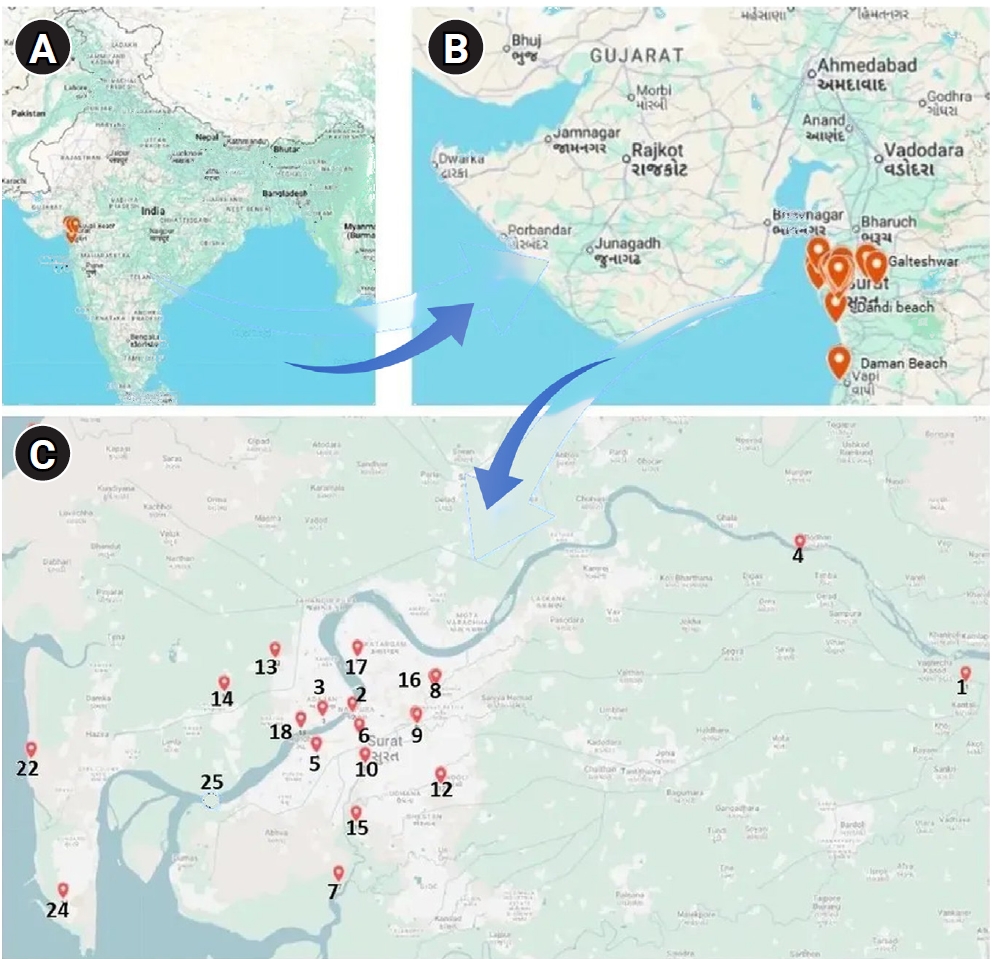
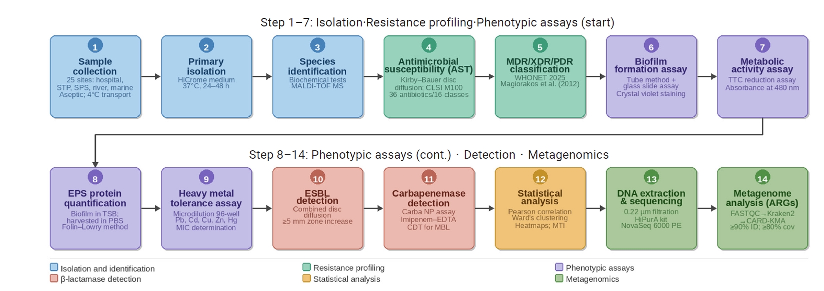
Figure 1.
Geographic location of the study area and sampling sites in Surat, Gujarat, India. The multi-panel map illustrates the sampling region at increasing spatial resolution. (A) Map of India highlighting Gujarat. (B) Regional map of the Gujarat coastline indicating Surat district. (C) Detailed spatial distribution of the 25 environmental sampling sites. Markers indicate specific collection points for river, sewage, and marine samples: 1, Bhamaiya, Tapi River; 2, Navdi Ovara, Tapi River; 3, Tapi River cable bridge; 4, Galteshwar, Tapi River; 5, Umra South sewage pumping station (SPS); 6, Khatodara SPS; 7, Khajod sewage treatment plant (STP); 8, Geetanagar STP; 9, Pratap Nagar STP; 10, Bhatar STP; 11, Anjana STP; 12, Dindoli STP; 13, Bhesan STP; 14, Aasarma STP; 15, Bamroli STP; 16, Karanj STP; 17, Siganpore STP; 18, Pal SPS; 19, SMIMER Hospital drainage; 20, Dandi beach coastal area; 21, Olpad beach; 22, Suvali beach; 23, Daman beach; 24, Hazira beach; and 25, Tapi River estuary.
Objectives: This study characterized multidrug-resistant (MDR) bacteria in sewage, river, and marine ecosystems in South Gujarat, India. Methods: Water samples were collected from 25 hospital drainage, sewage treatment/pumping, Tapi River, and coasta
Figure 2.
Multiphasic methodological workflow of the study. The sequential process included (1–3) sample isolation and MALDI-TOF MS identification; (4–5) resistance profiling and MDR classification; (6–11) phenotypic assays for biofilm formation, metabolic activity, heavy metal tolerance, and β-lactamase detection; (12) statistical analysis; and (13–14) NovaSeq 6000 sequencing and metagenomic identification of ARGs using the CARD-KMA pipeline. STP, sewage treatment plant; SPS, sewage pumping station; MALDI-TOF MS, matrix-assisted laser desorption/ionization time-of-flight mass spectrometry; AST, antimicrobial susceptibility testing; CLSI, Clinical and Laboratory Standards Institute; MDR, multidrug-resistant; XDR, extensively drug-resistant; PDR, pan-drug-resistant; TTC, 2,3,5-triphenyltetrazolium chloride; EPS, extracellular polymeric substance; TSB, tryptic soy broth; PBS, phosphate-buffered saline; Pb, lead; Cd, cadmium; Cu, copper; Zn, zinc; Hg, mercury; MIC, minimum inhibitory concentration; ESBL, extended-spectrum β-lactamase; EDTA, ethylenediaminetetraacetic acid; CDT, combined disc test; MBL, metallo-β-lactamase; MTI, metal tolerance index; ARGs, antibiotic resistance genes; CARD, Comprehensive Antibiotic Resistance Database; KMA, K-mer Alignment.
Objectives: This study characterized multidrug-resistant (MDR) bacteria in sewage, river, and marine ecosystems in South Gujarat, India. Methods: Water samples were collected from 25 hospital drainage, sewage treatment/pumping, Tapi River, and coasta
Figure 3.
Primary isolation of bacterial isolates on HiCrome Universal Differential Medium (HiMedia Laboratories Pvt. Ltd.), showing representative mixed colony morphologies and chromogenic differentiation used for presumptive grouping before further phenotypic and matrix-assisted laser desorption/ionization time-of-flight mass spectrometry–based identification.
Objectives: This study characterized multidrug-resistant (MDR) bacteria in sewage, river, and marine ecosystems in South Gujarat, India. Methods: Water samples were collected from 25 hospital drainage, sewage treatment/pumping, Tapi River, and coasta
Figure 4.
Distribution and frequency of (A) Gram-negative bacterial species and (B) Gram-positive bacterial species and unidentified isolates identified by matrix-assisted laser desorption/ionization time-of-flight mass spectrometry. Values represent the absolute number of isolates per species.
Objectives: This study characterized multidrug-resistant (MDR) bacteria in sewage, river, and marine ecosystems in South Gujarat, India. Methods: Water samples were collected from 25 hospital drainage, sewage treatment/pumping, Tapi River, and coasta
Figure 5.
Distribution of antimicrobial resistance phenotypes among bacterial isolates. The inner ring shows the overall prevalence of multidrug-resistant (MDR), extensively drug-resistant (XDR), and pan-drug-resistant (PDR) isolates, whereas the outer ring depicts their proportional distribution across sewage, marine, and river water sources.
Objectives: This study characterized multidrug-resistant (MDR) bacteria in sewage, river, and marine ecosystems in South Gujarat, India. Methods: Water samples were collected from 25 hospital drainage, sewage treatment/pumping, Tapi River, and coasta
Figure 6.
Comprehensive assessment of biofilm formation dynamics in multidrug-resistant isolates (n=166). (A) Distribution of biofilm-forming ability based on qualitative scoring percentages. (B–E) Phenotypic assessment of biofilm formation in representative isolates using tube and glass slide methods. (B) Strong biofilm producer (SAMR117 Bordetella pertussis), showing a thick (>2 mm) crystal violet–stained ring at the air–liquid interface and dense cellular aggregates. (C) Moderate biofilm producer (SAMR95 Pseudomonas aeruginosa), exhibiting an approximately 2-mm-thick biofilm ring with intermediate adherence. (D) Weak biofilm producer (SAMR125 Stenotrophomonas maltophilia), displaying a thin, approximately 1-mm ring and sparse cellular aggregation. (E) Non–biofilm-forming control (SAMR73 Micrococcus endophyticus), showing no visible crystal violet retention. (F) Scatter plot showing the relationship between metabolic activity, measured as optical density (OD) at 480 nm, and total bacterial biomass, measured as protein concentration (µg/mL), color-coded by biofilm-forming capacity (score 0–2); Pearson correlation analysis indicated no significant linear association.
Objectives: This study characterized multidrug-resistant (MDR) bacteria in sewage, river, and marine ecosystems in South Gujarat, India. Methods: Water samples were collected from 25 hospital drainage, sewage treatment/pumping, Tapi River, and coasta
Figure 7.
Hierarchical clustering and heatmap of standardized heavy metal tolerance indices (Z scores) among environmental bacterial isolates. Rows represent individual isolates (n=166), annotated by resistance category and environmental source, and columns represent the tested metals cadmium, zinc, copper, mercury, and lead. MDR, multidrug-resistant; XDR, extensively drug-resistant.
Objectives: This study characterized multidrug-resistant (MDR) bacteria in sewage, river, and marine ecosystems in South Gujarat, India. Methods: Water samples were collected from 25 hospital drainage, sewage treatment/pumping, Tapi River, and coasta
Figure 8.
Comparison of mean relative heavy metal tolerance indices between MDR and XDR isolates. Bars represent mean values, and error bars indicate standard deviations. Statistical significance was assessed at p<0.05. MDR, multidrug-resistant; XDR, extensively drug-resistant.
Objectives: This study characterized multidrug-resistant (MDR) bacteria in sewage, river, and marine ecosystems in South Gujarat, India. Methods: Water samples were collected from 25 hospital drainage, sewage treatment/pumping, Tapi River, and coasta
Figure 9.
Pearson correlation heatmaps illustrating relationships between heavy metal tolerance indices and physiological parameters in MDR and XDR bacterial isolates. The analysis compares variables across 4 environmental and resistance profiles: (A) marine MDR isolates (n=38), (B) river MDR isolates (n=23), (C) sewage MDR isolates (n=76), and (D) sewage XDR isolates (n=19). The color gradient represents the strength and direction of the Pearson correlation coefficient (r), ranging from dark blue (strong negative correlation, −1.0) to dark red (strong positive correlation, +1.0). Numerical values within each cell indicate the corresponding r value. Variables include tolerance indices for cadmium (Cd), lead (Pb), zinc (Zn), copper (Cu), and mercury (Hg), along with the CMTI, biofilm formation, metabolic OD, and protein content. Strong positive clustering is observed between Zn and Cu across MDR groups, whereas distinct correlation patterns emerge in the XDR sewage population. MDR, multidrug-resistant; XDR, extensively drug-resistant; CMTI, composite metal tolerance index; OD, optical density.
Objectives: This study characterized multidrug-resistant (MDR) bacteria in sewage, river, and marine ecosystems in South Gujarat, India. Methods: Water samples were collected from 25 hospital drainage, sewage treatment/pumping, Tapi River, and coasta
Figure 10.
Phenotypic detection of β-lactamase and carbapenemase production in Escherichia coli. (A) Combined disc diffusion assay for extended-spectrum β-lactamase production on Mueller-Hinton agar. (B) Carba NP assay showing a color change indicative of carbapenemase-mediated imipenem hydrolysis. (C) Screening of presumptive metallo-β-lactamase producers using the imipenem–ethylenediaminetetraacetic acid combined disc test on Mueller-Hinton agar.
Objectives: This study characterized multidrug-resistant (MDR) bacteria in sewage, river, and marine ecosystems in South Gujarat, India. Methods: Water samples were collected from 25 hospital drainage, sewage treatment/pumping, Tapi River, and coasta
Figure 11.
Hierarchical taxonomic distribution and relative abundance of bacterial taxa. The Krona multilayered pie chart illustrates the taxonomic composition of the microbial community and represents nested hierarchies from phylum-level assignments (inner circles) to species-level assignments (outermost ring). The community was predominantly composed of Pseudomonadota, driven by the abundance of Aeromonadaceae, Enterobacteriaceae, and Pseudomonadaceae. The arc length of each segment is proportional to the relative abundance of the corresponding taxon. Key pathogenic species, including A. caviae (31%), E. coli (17%), and K. pneumoniae (5%), constituted a substantial proportion of the identified community.
Objectives: This study characterized multidrug-resistant (MDR) bacteria in sewage, river, and marine ecosystems in South Gujarat, India. Methods: Water samples were collected from 25 hospital drainage, sewage treatment/pumping, Tapi River, and coasta
Figure 12.
Distribution of antimicrobial resistance genes and mechanisms. (A) Pie chart showing the overall proportions of resistance gene classes detected in the dataset, with efflux pumps (36.2%) and target-site mutations (21.3%) as the dominant mechanisms. (B) Stacked bar chart showing the number of antibiotic resistance genes (ARGs) per organism, color-coded by gene class; Escherichia coli and Acinetobacter baumannii exhibited the highest genetic diversity of resistance traits. (C) Sankey diagram linking ARG classes (left), specific genes (middle), and reported organisms (right). This visualization maps the distribution of resistance mechanisms and genetic determinants across bacterial taxa in the dataset.
Objectives: This study characterized multidrug-resistant (MDR) bacteria in sewage, river, and marine ecosystems in South Gujarat, India. Methods: Water samples were collected from 25 hospital drainage, sewage treatment/pumping, Tapi River, and coasta
Table 1.
Antibiotics used for antimicrobial susceptibility testing of bacterial isolates, including antibiotic class, subclass, standard abbreviation, and disc potency, according to Clinical and Laboratory Standards Institute guidelines
Table 1.
No. Antimicrobial class Subclass/Category Code Antimicrobial agent Disc abbreviation used Disc concentration
1 Penicillin Aminopenicillin (PEN) Ampicillin AMP 10 µg
Aminopenicillin+β-lactamase inhibitor (PEN+INH) Amoxicillin/clavulanic acid AMC 30 µg
Penicillinase-stable penicillin Oxacillin OX 1 µg
2 β-lactam/β-lactamase inhibitor combination β-lactam/β-lactamase inhibitor combination (BL+INH) Cefotaxime/clavulanic acid CEC 30/10 µg
Ceftazidime/clavulanic acid CAC 30/10 µg
Piperacillin/tazobactam PIT 100/10 µg
Ticarcillin/clavulanic acid TCC 75/10 µg
3 Cephalosporin First-generation cephalosporin (CEPH1) Cefazolin CZ 30 µg
Cephalothin CEP 30 µg
Second-generation cephalosporin (CEPH2) Cefuroxime CFM 5 µg
Third-generation cephalosporin (CEPH3) Cefotaxime CTX 30 µg
Ceftazidime CAZ 30 µg
Fourth-generation cephalosporin (CEPH4) Ceftriaxone CTR 30 µg
Cefepime CPM 30 µg
Cephamycin Cefoxitin CX 30 µg
4 Penems Carbapenem Imipenem IPM 10 µg
Ertapenem ETP 10 µg
Meropenem MRP 10 µg
5 Aminoglycoside Aminoglycoside Gentamicin GEN 10 µg
Netilmicin NET 30 µg
Amikacin AK 30 µg
Tobramycin TOB 10 µg
6 Quinolone Fluoroquinolones Ciprofloxacin CIP 30 µg
Ofloxacin OF 5 µg
Norfloxacin NX 10 µg
Pefloxacin PF 5 µg
Quinolone Nalidixic acid NA 30 µg
7 Nitroheterocyclics Nitroheterocyclics Nitrofurantoin NIT 300 µg
8 Folate pathway antagonists Folate pathway antagonist combination Cotrimoxazole COT 25 µg
9 Phenicol Phenicols Chloramphenicol C 30 µg
10 Tetracycline Tetracycline Minocycline MI 30 µg
Tetracycline TE 30 µg
Doxycycline hydrochloride DO 30 µg
Tetracycline/glycylcycline (GLY) Tigecycline TGC 15 µg
11 Macrolides Macrolide Erythromycin E 15 µg
Azithromycin AZM 15 µg
12 Oxazolidinone Oxazolidinone Linezolid LZ 30 µg
Lincosamides Lincosamide Clindamycin CD 2 µg
13 Fosfomycin Fosfomycin Fosfomycin FO 50 µg
14 Lipopeptides Polymyxins Colistin CS 10 µg
Polymyxin B PB 300 units
15 Monobactam Monobactam Aztreonam AT 30 µg
16 Glycopeptides Glycopeptides Vancomycin VA 30 µg
Lipoglycopeptides Teicoplanin TEI 30 µg
  • 1. Antimicrobial Resistance Collaborators. Global burden of bacterial antimicrobial resistance in 2019: a systematic analysis. Lancet 2022;399:629−55.
  • 2. Kariuki S. Global burden of antimicrobial resistance and forecasts to 2050. Lancet 2024;404:1172−3.
  • 3. EFSA Panel on Biological Hazards (BIOHAZ), Koutsoumanis K, Allende A, et al. Role played by the environment in the emergence and spread of antimicrobial resistance (AMR) through the food chain. EFSA J 2021;19:e06651.
  • 4. Gozlan RE. Understanding AM. In: Elumalai P, Lakshmi S, editors. Antimicrobial resistance in aquaculture and aquatic environments. Springer; 2025. p. 1−15.
  • 5. Singh B, Bhat A, Ravi K. Antibiotics misuse and antimicrobial resistance development in agriculture: a global challenge. Environ Health (Wash) 2024;2:618−22.
  • 6. Al-Khalaifah H, Rahman MH, Al-Surrayai T, et al. A One-Health perspective of antimicrobial resistance (AMR): human, animals and environmental health. Life (Basel) 2025;15:1598.
  • 7. Rizzo L, Manaia C, Merlin C, et al. Urban wastewater treatment plants as hotspots for antibiotic resistant bacteria and genes spread into the environment: a review. Sci Total Environ 2013;447:345−60.
  • 8. Baquero F, Coque TM, de la Cruz F. Ecology and evolution as targets: the need for novel eco-evo drugs and strategies to fight antibiotic resistance. Antimicrob Agents Chemother 2011;55:3649−60.
  • 9. Zielinski W, Korzeniewska E, Harnisz M, et al. Wastewater treatment plants as a reservoir of integrase and antibiotic resistance genes: an epidemiological threat to workers and environment. Environ Int 2021;156:106641.
  • 10. Wasko I, Kozinska A, Kotlarska E, et al. Clinically relevant b-lactam resistance genes in wastewater treatment plants. Int J Environ Res Public Health 2022;19:13829.
  • 11. Zhang S, Huang J, Zhao Z, et al. Hospital wastewater as a reservoir for antibiotic resistance genes: a meta-analysis. Front Public Health 2020;8:574968.
  • 12. Barathan M, Ng SL, Lokanathan Y, et al. Unseen weapons: bacterial extracellular vesicles and the spread of antibiotic resistance in aquatic environments. Int J Mol Sci 2024;25:3080.
  • 13. Pinto RM, Soares FA, Reis S, et al. Innovative strategies toward the disassembly of the EPS matrix in bacterial biofilms. Front Microbiol 2020;11:952.
  • 14. Singh S, Datta S, Narayanan KB, et al. Bacterial exo-polysaccharides in biofilms: role in antimicrobial resistance and treatments. J Genet Eng Biotechnol 2021;19:140.
  • 15. Hanna N, Tamhankar AJ, Stalsby Lundborg C. Antibiotic concentrations and antibiotic resistance in aquatic environments of the WHO Western Pacific and South-East Asia regions: a systematic review and probabilistic environmental hazard assessment. Lancet Planet Health 2023;7:e45−54.
  • 16. Taneja N, Sharma M. Antimicrobial resistance in the environment: the Indian scenario. Indian J Med Res 2019;149:119−28.
  • 17. Mohamed Hatha AA, Divya PS, Reshma S, et al. Polluted coastal and estuarine environments: a potential reservoir for AMR determinants in various pathogenic bacteria. In: Thomas S, editor. Antimicrobial resistance. Springer; 2020. p. 101−34.
  • 18. Goh SG, You L, Ng C, et al. A multi-pronged approach to assessing antimicrobial resistance risks in coastal waters and aquaculture systems. Water Res 2024;266:122353.
  • 19. Ram B, Kumar M. Correlation appraisal of antibiotic resistance with fecal, metal and microplastic contamination in a tropical Indian river, lakes and sewage. NPJ Clean Water 2020;3:3.
  • 20. Baker-Austin C, Wright MS, Stepanauskas R, et al. Co-selection of antibiotic and metal resistance. Trends Microbiol 2006;14:176−82.
  • 21. Seiler C, Berendonk TU. Heavy metal driven co-selection of antibiotic resistance in soil and water bodies impacted by agriculture and aquaculture. Front Microbiol 2012;3:399.
  • 22. Doi Y, Park YS, Rivera JI, et al. Community-associated extended-spectrum β-lactamase-producing Escherichia coli infection in the United States. Clin Infect Dis 2013;56:641−8.
  • 23. World Health Organization (WHO) (CH). WHO bacterial priority pathogens list, 2024: bacterial pathogens of public health importance, to guide research, development and strategies to prevent and control antimicrobial resistance. WHO; 2024.
  • 24. Leonard AFC, Zhang L, Balfour AJ, et al. Exposure to and colonisation by antibiotic-resistant E. coli in UK coastal water users: environmental surveillance, exposure assessment, and epidemiological study (Beach Bum Survey). Environ Int 2018;114:326−33.
  • 25. Bevan ER, Jones AM, Hawkey PM. Global epidemiology of CTX-M b-lactamases: temporal and geographical shifts in genotype. J Antimicrob Chemother 2017;72:2145−55.
  • 26. Algammal AM, Behzadi P. Antimicrobial resistance: a global public health concern that needs perspective combating strategies and new talented antibiotics. Discov Med 2024;36:1911−3.
  • 27. Karampatakis T, Tsergouli K, Behzadi P. Carbapenem-resistant Acinetobacter baumannii: virulence factors, molecular epidemiology, and latest updates in treatment options. Microorganisms 2025;13:1983.
  • 28. Karampatakis T, Tsergouli K, Behzadi P. Pan-genome plasticity and virulence factors: a natural treasure trove for Acinetobacter baumannii. Antibiotics (Basel) 2024;13:257.
  • 29. Ahmadi S, Foroohi F, Shirzadian M, et al. Distribution of acquired carbapenemase genes among uropathogenic Escherichia coli. Naunyn Schmiedebergs Arch Pharmacol 2026;399:3477−94.
  • 30. Kebriaee AN, Behzadi P, Mohammadian T, et al. Distribution of antimicrobial resistance genes and virulence genes in different genotypes of Acinetobacter baumannii. BMC Infect Dis 2025;25:1230.
  • 31. Karampatakis T, Tsergouli K, Behzadi P. Carbapenem-resistant Pseudomonas aeruginosa’s resistome: pan-genomic plasticity, the impact of transposable elements and jumping genes. Antibiotics (Basel) 2025;14:353.
  • 32. MacFaddin JF. Biochemical tests for identification of medical bacteria. 3rd ed. Lippincott Williams & Wilkins; 2000.
  • 33. Singhal N, Kumar M, Kanaujia PK, et al. MALDI-TOF mass spectrometry: an emerging technology for microbial identification and diagnosis. Front Microbiol 2015;6:791.
  • 34. Hudzicki J. Kirby-Bauer disk diffusion susceptibility test protocol [Internet]. American Society for Microbiology; 2009 [cited 2026 Jun 26]. Available from: https://asm.org/getattachment/2594ce26-bd44-47f6-8287-0657aa9185ad/kirby-bauer-disc-diffusion-susceptibility-test-protocol-pdf.pdf.
  • 35. Teshome MS, Meskel DH, Wondafrash B. Determinants of anemia among pregnant women attending antenatal care clinic at public health facilities in Kacha Birra District, Southern Ethiopia. J Multidiscip Healthc 2020;13:1007−15.
  • 36. Vansia A, Patel R, Dudhagara P. Analysis of multidrug resistance profile of Escherichia coli from clinical samples from companion animals and bird retrospect to five-year (2015-2019) literature data. Biointerface Res Appl Chem 2021;11:12506−15.
  • 37. Clinical and Laboratory Standards Institute (CLSI) (US). M100 Performance standards for antimicrobial susceptibility testing. CLSI; 2023.
  • 38. Clinical and Laboratory Standards Institute (CLSI) (US). M100 Performance standards for antimicrobial susceptibility testing. CLSI; 2024.
  • 39. Clinical and Laboratory Standards Institute (CLSI) (US). M100 Performance standards for antimicrobial susceptibility testing. CLSI; 2025.
  • 40. Magiorakos AP, Srinivasan A, Carey RB, et al. Multidrug-resistant, extensively drug-resistant and pandrug-resistant bacteria: an international expert proposal for interim standard definitions for acquired resistance. Clin Microbiol Infect 2012;18:268−81.
  • 41. Harika K, Shenoy VP, Narasimhaswamy N, et al. Detection of biofilm production and its impact on antibiotic resistance profile of bacterial isolates from chronic wound infections. J Glob Infect Dis 2020;12:129−34.
  • 42. Coraça-Huber DC, Kreidl L, Steixner S, et al. Identification and morphological characterization of biofilms formed by strains causing infection in orthopedic implants. Pathogens 2020;9:649.
  • 43. Jiao Y, Cody GD, Harding AK, et al. Characterization of extracellular polymeric substances from acidophilic microbial biofilms. Appl Environ Microbiol 2010;76:2916−22.
  • 44. Lowry OH, Rosebrough NJ, Farr AL, et al. Protein measurement with the Folin phenol reagent. J Biol Chem 1951;193:265−75.
  • 45. DiVecchia L, Somerson NL. Tetrazolium reduction as a measure of metabolic activity for glass-adherent Mycoplasma pneumoniae. Appl Microbiol 1973;26:298−302.
  • 46. Pagnucco G, Overfield D, Chamlee Y, et al. Metal tolerance and biosorption capacities of bacterial strains isolated from an urban watershed. Front Microbiol 2023;14:1278886.
  • 47. Ojo GJ, Onile OS, Momoh AO, et al. Physiochemical analyses and molecular characterization of heavy metal-resistant bacteria from Ilesha gold mining sites in Nigeria. J Genet Eng Biotechnol 2023;21:172.
  • 48. Nordmann P, Naas T, Poirel L. Global spread of carbapenemase-producing Enterobacteriaceae. Emerg Infect Dis 2011;17:1791−8.
  • 49. Vamsi KS, Moorthy SR, Murali TS, et al. Phenotypic methods for the detection of metallo-beta-lactamase production by gram-negative bacterial isolates from hospitalized patients in a tertiary care hospital in India. J Pure Appl Microbiol 2021;15:2019−26.
  • 50. Andrews S. FastQC: a quality control tool for high throughput sequence data [Internet]. Babraham Bioinformatics; 2010 [cited 2026 Jun 26]. Available from: https://www.bioinformatics.babraham.ac.uk/projects/fastqc/.
  • 51. Wood DE, Lu J, Langmead B. Improved metagenomic analysis with Kraken 2. Genome Biol 2019;20:257.
  • 52. Ondov BD, Bergman NH, Phillippy AM. Interactive metagenomic visualization in a Web browser. BMC Bioinformatics 2011;12:385.
  • 53. Alcock BP, Huynh W, Chalil R, et al. CARD 2023: expanded curation, support for machine learning, and resistome prediction at the Comprehensive Antibiotic Resistance Database. Nucleic Acids Res 2023;51:D690−9.
  • 54. Clausen PT, Aarestrup FM, Lund O. Rapid and precise alignment of raw reads against redundant databases with KMA. BMC Bioinformatics 2018;19:307.
  • 55. Olson RD, Assaf R, Brettin T, et al. Introducing the Bacterial and Viral Bioinformatics Resource Center (BV-BRC): a resource combining PATRIC, IRD and ViPR. Nucleic Acids Res 2023;51:D678−89.
  • 56. Kummerer K. Antibiotics in the aquatic environment: a review--part II. Chemosphere 2009;75:435−41.
  • 57. Tacao M, Correia A, Henriques IS. Low prevalence of carbapenem-resistant bacteria in river water: resistance is mostly related to intrinsic mechanisms. Microb Drug Resist 2015;21:497−506.
  • 58. Baquero F, Martinez JL, Canton R. Antibiotics and antibiotic resistance in water environments. Curr Opin Biotechnol 2008;19:260−5.
  • 59. Wellington EM, Boxall AB, Cross P, et al. The role of the natural environment in the emergence of antibiotic resistance in gram-negative bacteria. Lancet Infect Dis 2013;13:155−65.
  • 60. Manaia CM. Assessing the risk of antibiotic resistance transmission from the environment to humans: non-direct proportionality between abundance and risk. Trends Microbiol 2017;25:173−81.
  • 61. Carvalho IT, Santos L. Antibiotics in the aquatic environments: a review of the European scenario. Environ Int 2016;94:736−57.
  • 62. Saini S, Tewari S, Dwivedi J, et al. Biofilm-mediated wastewater treatment: a comprehensive review. Mater Adv 2023;4:1415−43.
  • 63. Poli A, Anzelmo G, Nicolaus B. Bacterial exopolysaccharides from extreme marine habitats: production, characterization and biological activities. Mar Drugs 2010;8:1779−802.
  • 64. Lebeaux D, Ghigo JM, Beloin C. Biofilm-related infections: bridging the gap between clinical management and fundamental aspects of recalcitrance toward antibiotics. Microbiol Mol Biol Rev 2014;78:510−43.
  • 65. Mah TF, O'Toole GA. Mechanisms of biofilm resistance to antimicrobial agents. Trends Microbiol 2001;9:34−9.
  • 66. Flemming HC, Wuertz S. Bacteria and archaea on Earth and their abundance in biofilms. Nat Rev Microbiol 2019;17:247−60.
  • 67. Ducret V, Gonzalez MR, Leoni S, et al. The CzcCBA Efflux system requires the CadA P-type ATPase for timely expression upon zinc excess in Pseudomonas aeruginosa. Front Microbiol 2020;11:911.
  • 68. Chua SL, Yam JK, Hao P, et al. Selective labelling and eradication of antibiotic-tolerant bacterial populations in Pseudomonas aeruginosa biofilms. Nat Commun 2016;7:10750.
  • 69. Valentini M, Filloux A. Biofilms and cyclic di-GMP (c-di-GMP) signaling: lessons from Pseudomonas aeruginosa and other bacteria. J Biol Chem 2016;291:12547−55.
  • 70. Teitzel GM, Parsek MR. Heavy metal resistance of biofilm and planktonic Pseudomonas aeruginosa. Appl Environ Microbiol 2003;69:2313−20.
  • 71. Boolchandani M, D'Souza AW, Dantas G. Sequencing-based methods and resources to study antimicrobial resistance. Nat Rev Genet 2019;20:356−70.
  • 72. Tamma PD, Aitken SL, Bonomo RA, et al. Infectious Diseases Society of America Guidance on the treatment of extended-spectrum β-lactamase producing Enterobacterales (ESBL-E), carbapenem-resistant Enterobacterales (CRE), and Pseudomonas aeruginosa with difficult-to-treat resistance (DTR- P. aeruginosa). Clin Infect Dis 2021;72:e169−83.
  • 73. Marti E, Variatza E, Balcazar JL. The role of aquatic ecosystems as reservoirs of antibiotic resistance. Trends Microbiol 2014;22:36−41.

Download Citation

Download a citation file in RIS format that can be imported by all major citation management software, including EndNote, ProCite, RefWorks, and Reference Manager.

Format:

Include:

Environmental emergence and dissemination of clinically relevant multidrug-resistant bacteria and resistance genes in sewage and aquatic ecosystems
Download Citation

Download a citation file in RIS format that can be imported by all major citation management software, including EndNote, ProCite, RefWorks, and Reference Manager.

Format:
Include:
Environmental emergence and dissemination of clinically relevant multidrug-resistant bacteria and resistance genes in sewage and aquatic ecosystems
Close

Figure